Copyright
©The Author(s) 2022.
World J Clin Cases. Sep 26, 2022; 10(27): 9936-9944
Published online Sep 26, 2022. doi: 10.12998/wjcc.v10.i27.9936
Published online Sep 26, 2022. doi: 10.12998/wjcc.v10.i27.9936
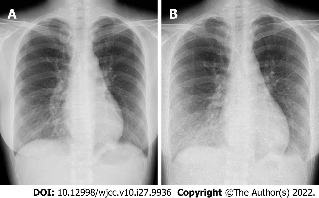
Figure 1 Chest radiography images.
A: Chest radiography at admission shows right lower lung interstitial shadows; B: Chest radiography on day 4 of hospitalization shows worsened right lower lung interstitial shadows.
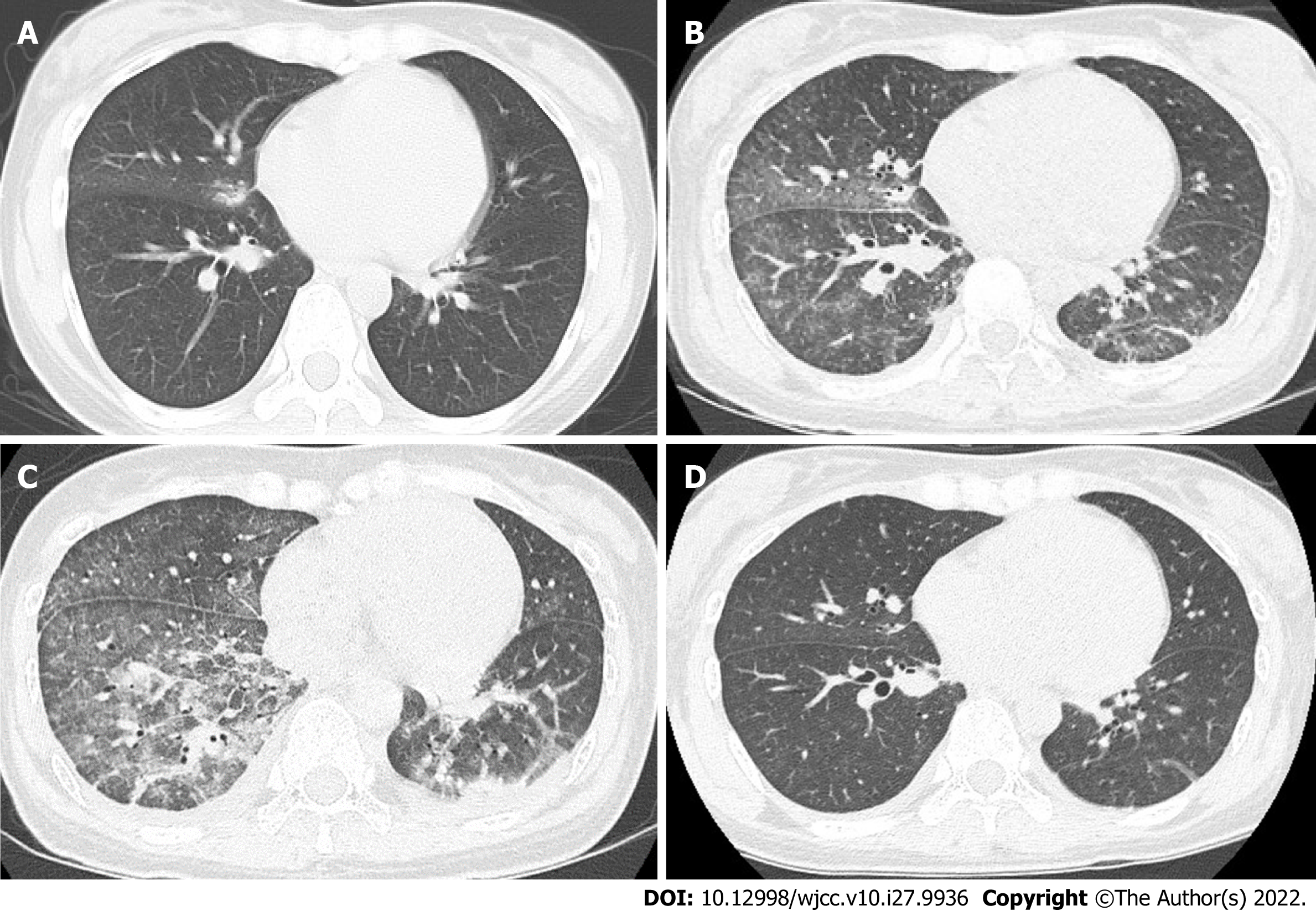
Figure 2 Chest computed tomography images.
A: Chest computed tomography at admission reveals right middle and lower lobar consolidation and ground-glass opacity; B and C: Chest computed tomography on day 4 of hospitalization shows extensive ground-glass opacity, mainly in the right middle and lower lobes, and thick bilateral basal infiltrative shadows. In addition, thickening of the interlobular septum and bronchovascular bundles, and bilateral pleural effusion, suggestive of eosinophilic or organizing pneumonia, are seen; D: Chest computed tomography on day 10 of hospitalization shows improvement of ground-glass opacity and infiltrative shadows.
- Citation: Fujii M, Kenzaka T. Drug-induced lung injury caused by acetaminophen in a Japanese woman: A case report. World J Clin Cases 2022; 10(27): 9936-9944
- URL: https://www.wjgnet.com/2307-8960/full/v10/i27/9936.htm
- DOI: https://dx.doi.org/10.12998/wjcc.v10.i27.9936


