Copyright
©The Author(s) 2022.
World J Clin Cases. Sep 26, 2022; 10(27): 9911-9920
Published online Sep 26, 2022. doi: 10.12998/wjcc.v10.i27.9911
Published online Sep 26, 2022. doi: 10.12998/wjcc.v10.i27.9911
Figure 1 Enhanced computerized tomography performed after the patient vomited approximately 500 mL of fresh blood and entered a state of hemorrhagic shock.
A: Cross section. The mid thoracic esophagus is dilated, and the esophageal lumen is filled with massive hematomas; B: Cross section. An occupying lesion with a relatively clear boundary is observed under the mucosa just above the esophagogastric (EG) junction, with partial contrast effects (orange arrow); C: Coronal section. An occupying lesion with a relatively clear boundary is observed under the mucosa just above the EG junction, with partial contrast effects (orange arrow).
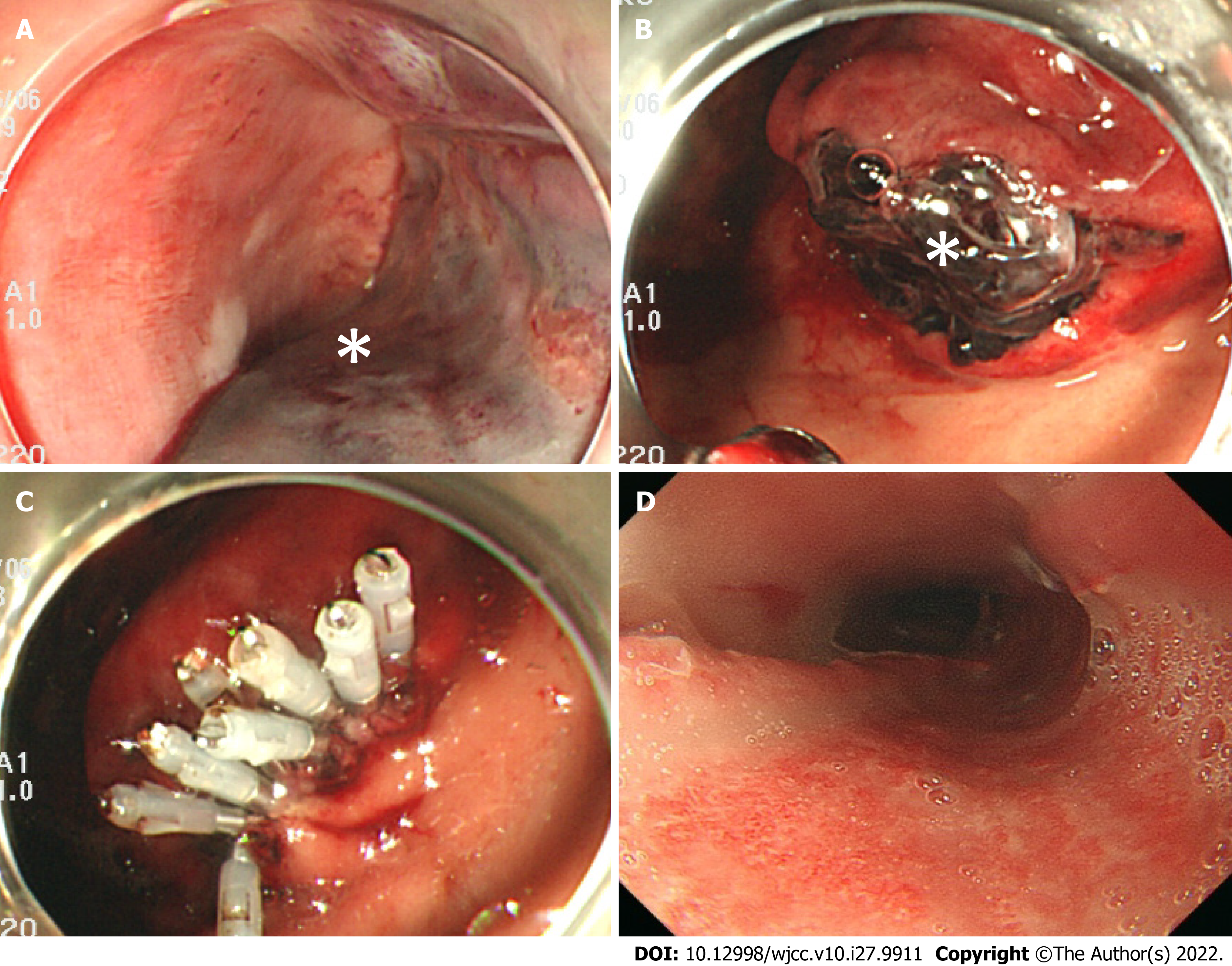
Figure 2 Upper gastrointestinal endoscopy images after hematemesis.
A: Middle esophagus. A longitudinal extension of reddish mucosal thickening (white asterisk) and obstructing of the esophagus are confirmed; B: Middle esophagus, temporary compression hemostasis was performed with Sengstaken-Blakemore tube (white asterisk); C: Lower esophagus, massive hematoma and laceration of gastric mucosa together with bleeding are confirmed in the esophagogastric junction (white asterisk);.
Figure 3 Upper gastrointestinal endoscopy images of postoperative course.
A: One day after surgery, middle esophagus, hemostasis is confirmed; B: One day after surgery, Lower esophagus, Hematoma (white asterisk) and laceration of the gastric mucosa are confirmed in the esophagogastric (EG) junction; C: One day after surgery, EG junction, gastric mucosal laceration is clipped; D: 10 d after surgery, middle esophagus, the submucosal hematoma has been replaced by an esophageal ulcer.
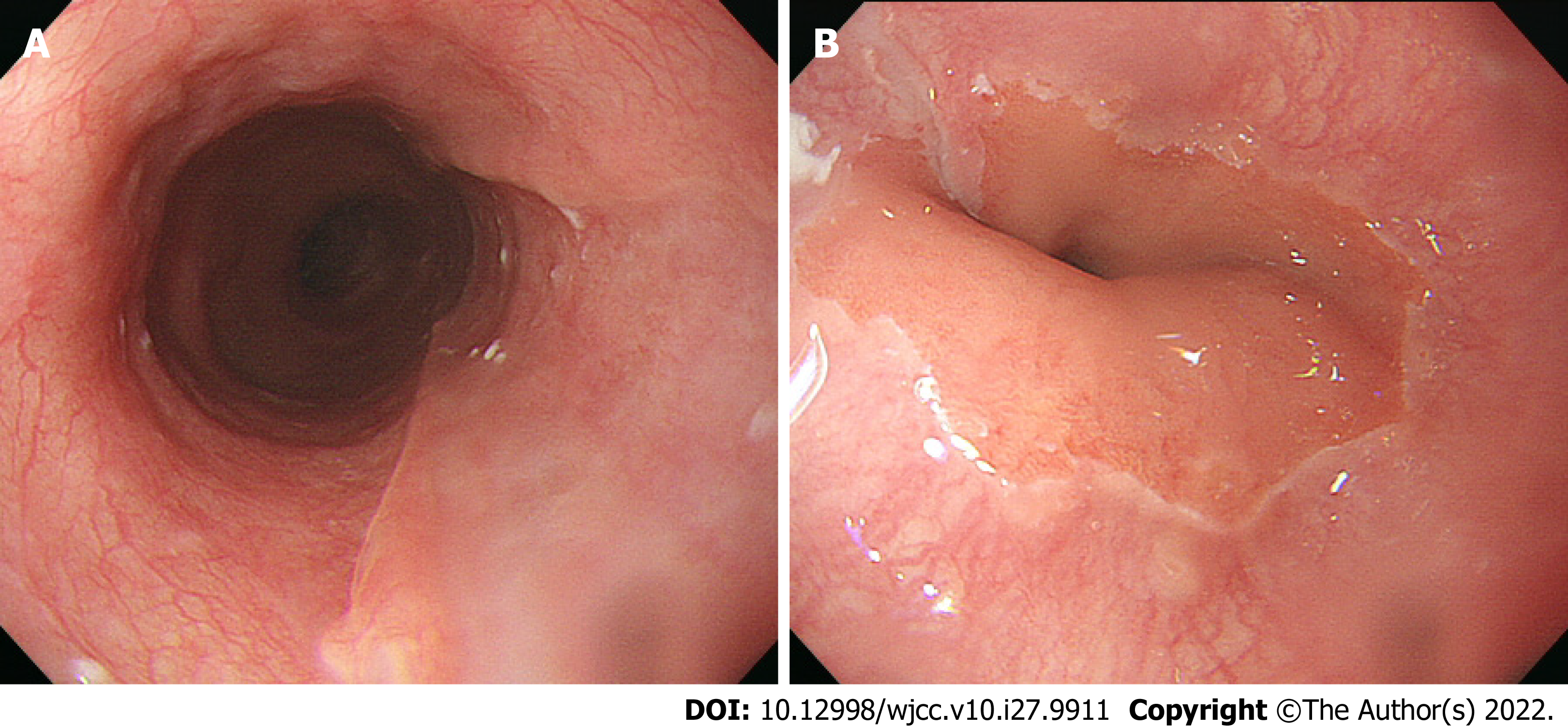
Figure 4 Upper gastrointestinal endoscopy images 60 d after surgery.
A: Middle esophagus, hemostasis has disappeared, and the endoscopy revealed normal findings; B: Esophagogastric junction, the esophageal ulcer has been replaced with a scar.
- Citation: Oba J, Usuda D, Tsuge S, Sakurai R, Kawai K, Matsubara S, Tanaka R, Suzuki M, Takano H, Shimozawa S, Hotchi Y, Usami K, Tokunaga S, Osugi I, Katou R, Ito S, Mishima K, Kondo A, Mizuno K, Takami H, Komatsu T, Nomura T, Sugita M. Hemorrhagic shock due to submucosal esophageal hematoma along with mallory-weiss syndrome: A case report. World J Clin Cases 2022; 10(27): 9911-9920
- URL: https://www.wjgnet.com/2307-8960/full/v10/i27/9911.htm
- DOI: https://dx.doi.org/10.12998/wjcc.v10.i27.9911


