Copyright
©The Author(s) 2022.
World J Clin Cases. Sep 26, 2022; 10(27): 9897-9903
Published online Sep 26, 2022. doi: 10.12998/wjcc.v10.i27.9897
Published online Sep 26, 2022. doi: 10.12998/wjcc.v10.i27.9897
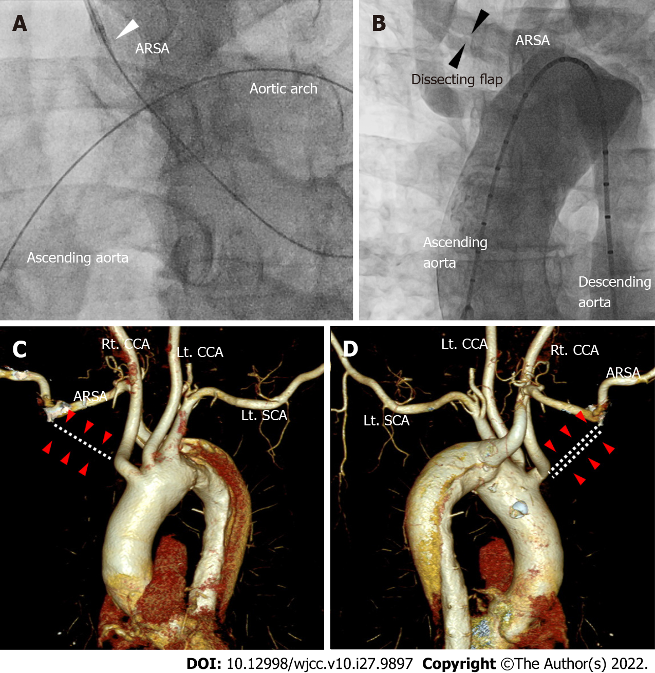
Figure 1 Aortography and computed tomography of aorta and aberrant right subclavian artery.
A: The guidewire reached the ascending aorta after forming a large loop in the left anterior oblique 30° view of coronary angiography. We were unable to advance the guiding catheter past the ostium of the right subclavian artery (white arrowhead); B: The aortogram using a 5Fr pigtail catheter via the right femoral artery shows a dissection flap of the right subclavian artery in the AP view (black arrowheads); C and D: The right subclavian artery did not originate from the right innominate artery (white dotted lines and red arrow heads in C and D). Instead, aberrant right subclavian artery emerged from the descending aorta. ARSA: Aberrant right subclavian artery; SCA: Subclavian artery; CCA: Common carotid artery.
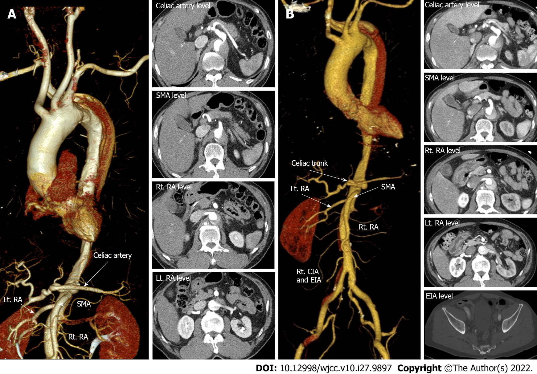
Figure 2 Initial computed tomography post transradial intervention and follow-up computed tomography in 2 days.
A: Emergency computed tomography (CT) aortography immediately after right transradial intervention showing type B aortic dissection (AD) originating from the aberrant right subclavian artery with extension of the intimal flap down the descending to the infrarenal abdominal aorta. The dissection extended into the left renal artery (RA) (red arrowheads in A); B: After two days of intensive care unit stay, follow-up CT showed downstream propagation of the AD into the external iliac artery and left RA with compromised flow (red arrowheads of B). Other arteries including the celiac trunk, superior mesenteric artery and right RA were intact (white arrows) on both CTs. SMA: Superior mesenteric artery; RA: Renal artery; EIA: External iliac artery; CIA: Common iliac artery.
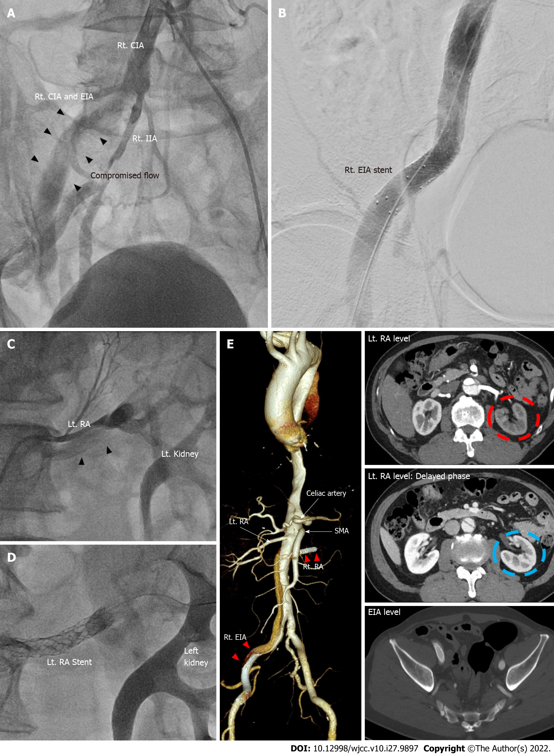
Figure 3 Percutaneous angioplasty of right common iliac artery and left renal artery and follow-up computed tomography in 8 mo.
A: Anteroposterior view of pre-intervention angiography showing sluggish blood flow in the right common iliac artery and external iliac artery (black arrowheads of A); B: Which was salvaged by stent implantation; C and D: Compromised blood flow of left renal artery pre-intervention (black arrowheads in C) was also recovered after by angioplasty (D); E: Follow-up computed tomography at eight months post intervention demonstrated patent stents without further propagation of aortic dissection. Left kidney perfusion was slightly delayed (red dotted circle of right upper panel of E) but preserved (blue dotted circle in the right middle panel of E). SMA: Superior mesenteric artery; RA: Renal artery; EIA: External iliac artery; CIA: Common iliac artery.
- Citation: Ha K, Jang AY, Shin YH, Lee J, Seo J, Lee SI, Kang WC, Suh SY. Iatrogenic aortic dissection during right transradial intervention in a patient with aberrant right subclavian artery: A case report. World J Clin Cases 2022; 10(27): 9897-9903
- URL: https://www.wjgnet.com/2307-8960/full/v10/i27/9897.htm
- DOI: https://dx.doi.org/10.12998/wjcc.v10.i27.9897


