Copyright
©The Author(s) 2022.
World J Clin Cases. Sep 26, 2022; 10(27): 9886-9896
Published online Sep 26, 2022. doi: 10.12998/wjcc.v10.i27.9886
Published online Sep 26, 2022. doi: 10.12998/wjcc.v10.i27.9886
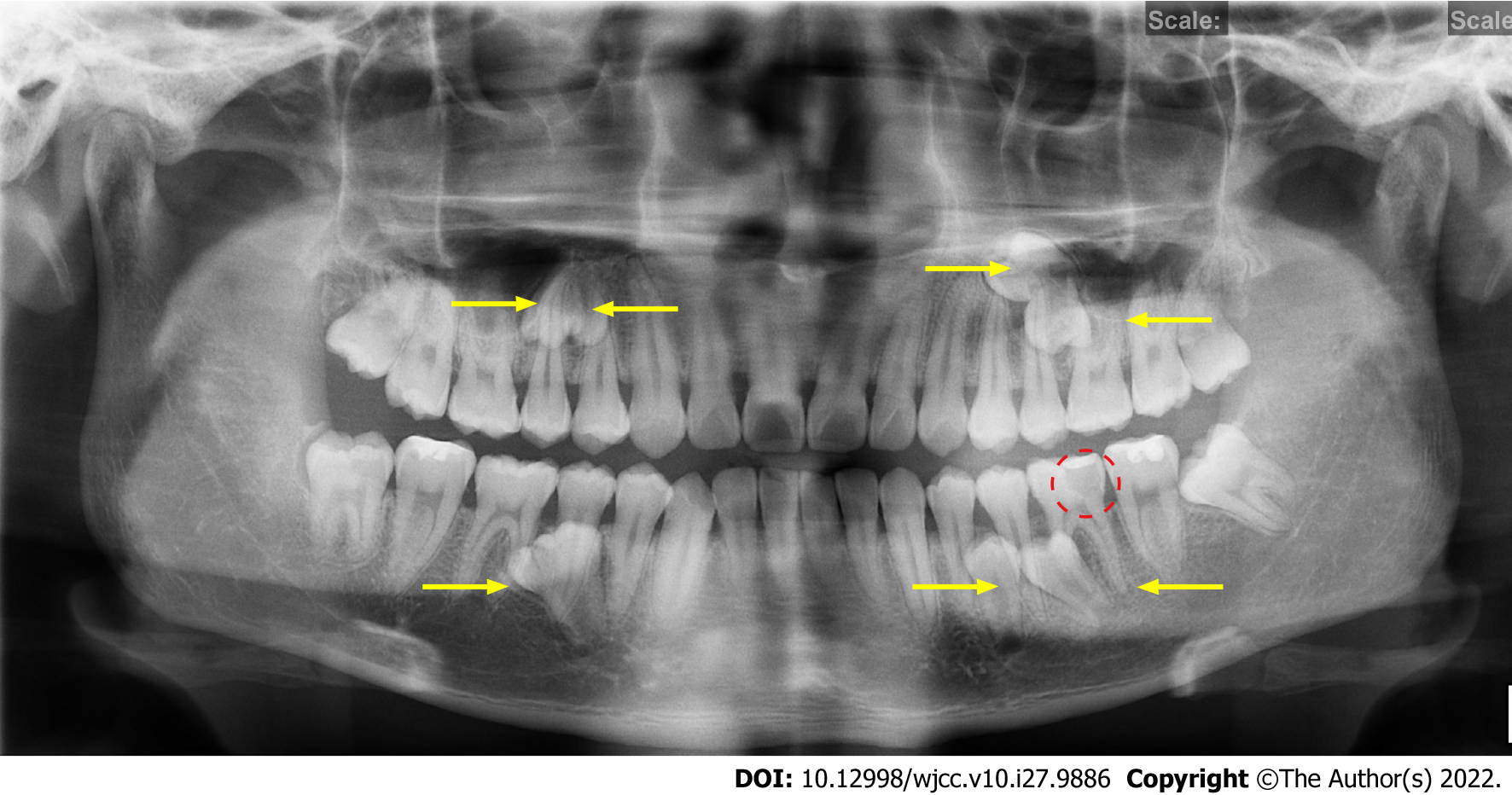
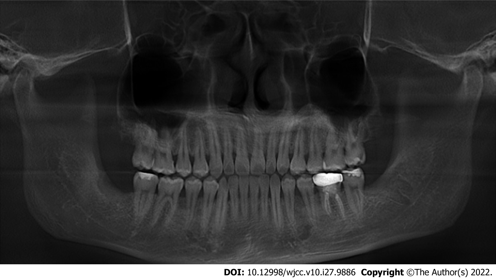
Figure 1 Preoperative panoramic radiograph.
The yellow arrows show the positions of the seven impacted SNTs. The red circle shows a local low-density area close to the pulp cavity in tooth #36.
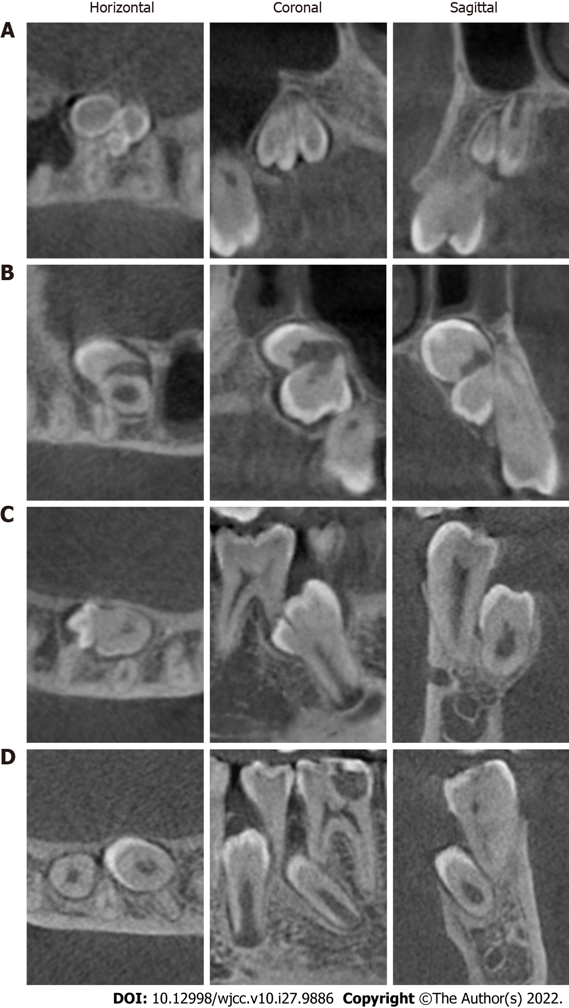
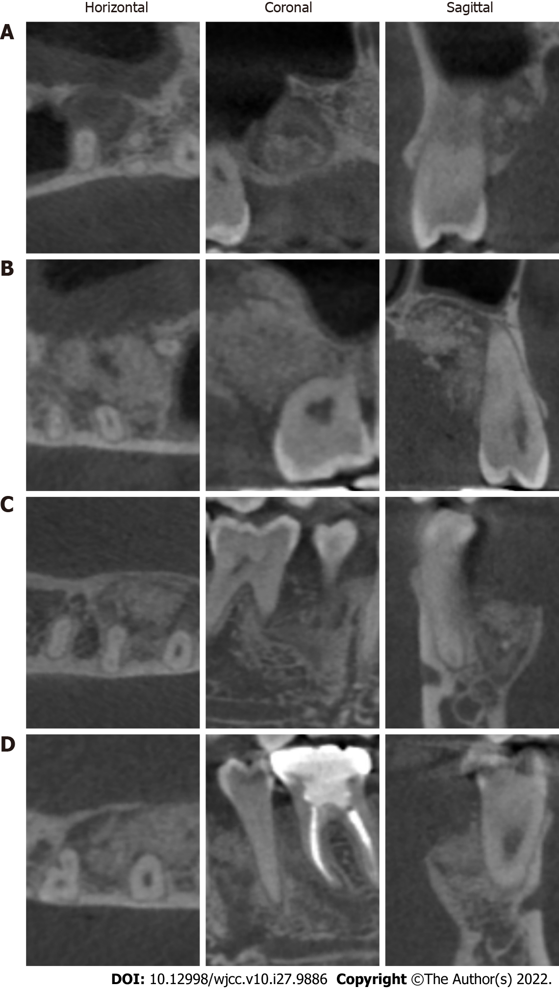
Figure 2 Preoperative cone-beam computed tomography images.
A: Right maxilla imaging of the supernumerary teeth (SNTs); B: Left maxilla imaging of the SNTs; C: Right mandible imaging of the SNTs; D: Left mandible imaging of the SNTs. Left: Horizontal images; Middle: Coronal images; Right: Sagittal images.
Figure 3 Treatment process for tooth #36.
A: Gutta-Percha cone fitting and working length confirmation with a periapical radiograph; B: Postobturation periapical radiograph. C: Restoration of tooth #36 with an all-ceramic crown.
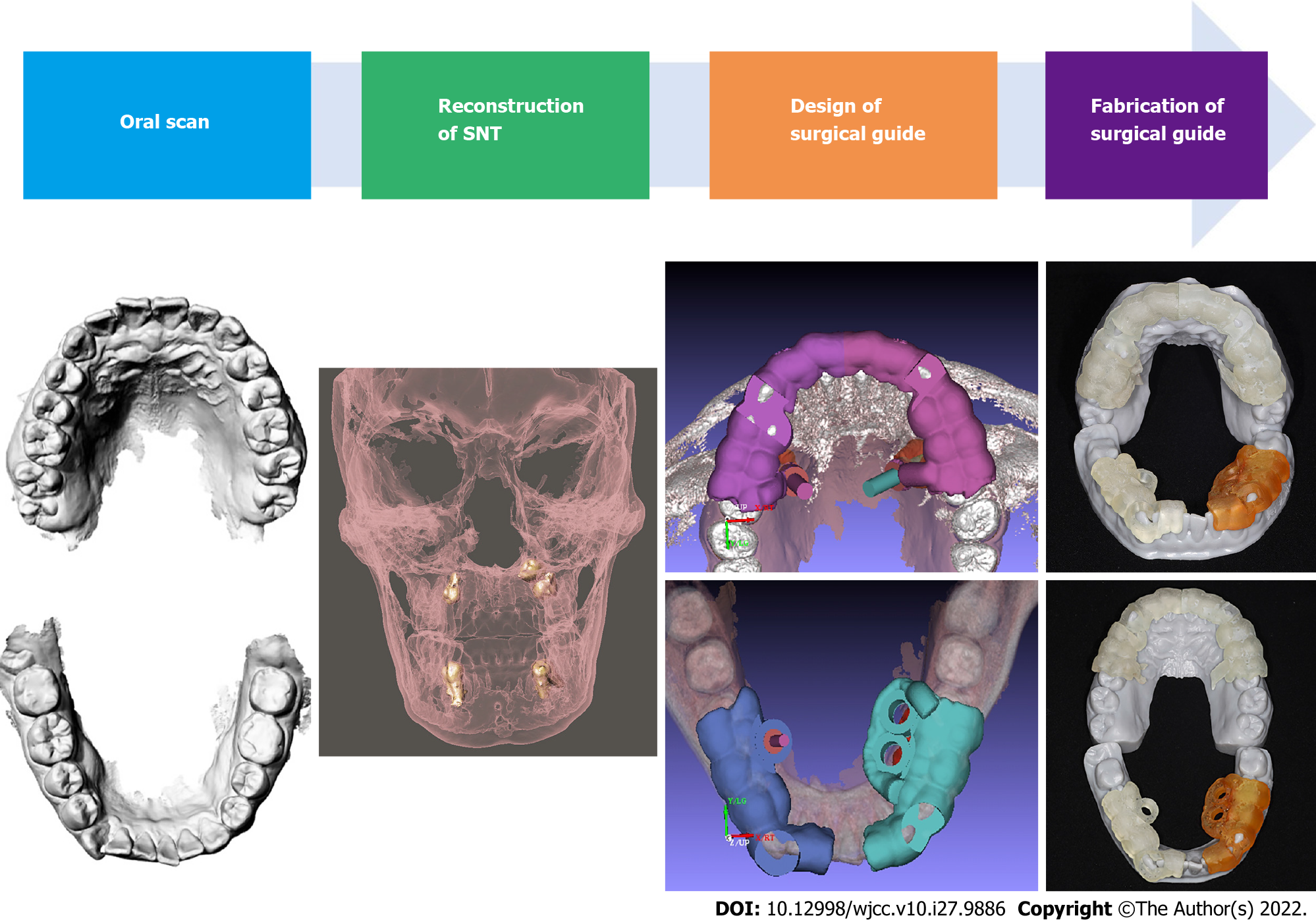
Figure 4 Design and fabrication process of the digital positioning guide plates.
From left to right, (1) oral scan; (2) reconstruction of the shapes of the impacted Supernumerary teeth; (3) preoperative analysis and design of the surgical guide plate with computer-aided design software; and (4) fabrication of the surgical guide plate with a (computer-aided manufacturing) 3D printer.
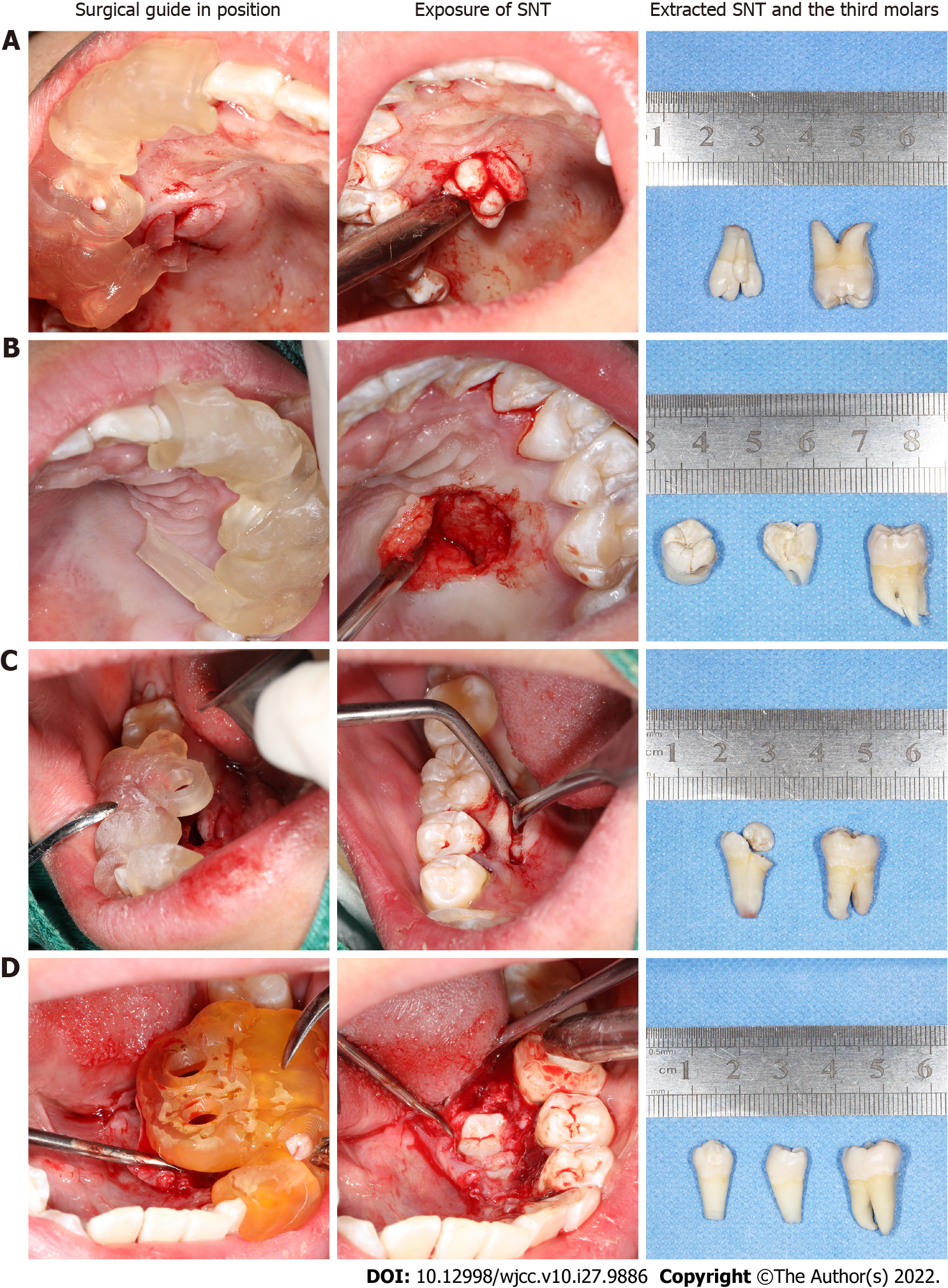
Figure 5 Surgical supernumerary teeth extraction procedure.
A: Supernumerary teeth (SNT) extraction from the right maxilla; B: SNT extraction from the left maxilla; C: SNT extraction from the right mandible; D: SNT extraction from the left mandible; from left to right: the surgical guide plate in position, exposure of the SNTs, and extracted SNTs and third molars.
Figure 6 Postoperative panoramic radiograph.
The seven impacted supernumerary teeth and two impacted mandibular third molars were completely extracted.
Figure 7 Postoperative cone-beam computed tomography imaging.
A: Right maxilla imaging of the supernumerary teeth (SNTs); B: Left maxilla imaging of the SNTs; C: Right mandible imaging of the SNTs; D: Left mandible imaging of the SNTs. Left: Horizontal images; Middle: Coronal images; Right: Sagittal images.
- Citation: Wang Z, Zhao SY, He WS, Yu F, Shi SJ, Xia XL, Luo XX, Xiao YH. Application of digital positioning guide plates for the surgical extraction of multiple impacted supernumerary teeth: A case report and review of literature. World J Clin Cases 2022; 10(27): 9886-9896
- URL: https://www.wjgnet.com/2307-8960/full/v10/i27/9886.htm
- DOI: https://dx.doi.org/10.12998/wjcc.v10.i27.9886


