Copyright
©The Author(s) 2022.
World J Clin Cases. Sep 26, 2022; 10(27): 9879-9885
Published online Sep 26, 2022. doi: 10.12998/wjcc.v10.i27.9879
Published online Sep 26, 2022. doi: 10.12998/wjcc.v10.i27.9879
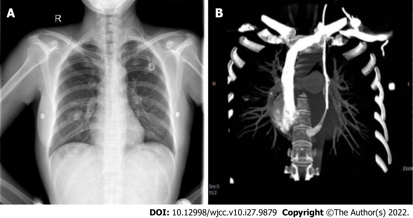
Figure 1 Orthotopic chest radiography after the operation.
A: The catheter walks beside the left mediastinum; B: The contrast medium flowed into the right atrium through the coronary sinus.
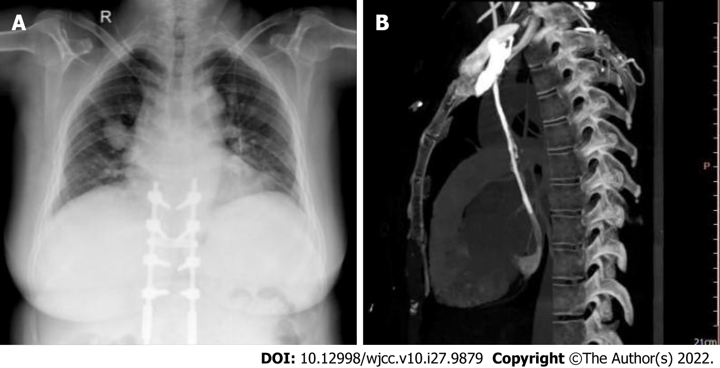
Figure 2 A chest X-ray examination after operation.
A: The catheter walks beside the left mediastinum; B: Persistent left superior vena cava converged into the right atrium through the coronary sinus.
- Citation: Zhou RN, Ma XB, Wang L, Kang HF. Accidental venous port placement via the persistent left superior vena cava: Two case reports. World J Clin Cases 2022; 10(27): 9879-9885
- URL: https://www.wjgnet.com/2307-8960/full/v10/i27/9879.htm
- DOI: https://dx.doi.org/10.12998/wjcc.v10.i27.9879


