Copyright
©The Author(s) 2022.
World J Clin Cases. Sep 26, 2022; 10(27): 9865-9872
Published online Sep 26, 2022. doi: 10.12998/wjcc.v10.i27.9865
Published online Sep 26, 2022. doi: 10.12998/wjcc.v10.i27.9865
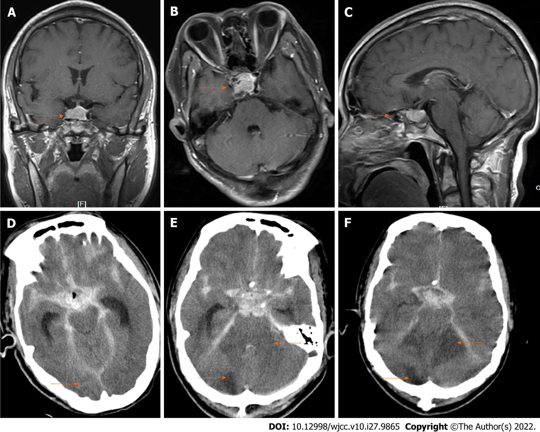
Figure 1 Preoperative magnetic resonance imaging and postoperative computed tomography images in case 1 (orange arrow).
A-C: Magnetic resonance imaging showed occupation in the sellar region; D-F: Computed tomography showed intracerebral hemorrhage on the day of surgery and progressive cerebral infarction on postoperative days 2 and 4, respectively.
- Citation: Wang J, Peng YM. Emergency treatment and anesthesia management of internal carotid artery injury during neurosurgery: Four case reports. World J Clin Cases 2022; 10(27): 9865-9872
- URL: https://www.wjgnet.com/2307-8960/full/v10/i27/9865.htm
- DOI: https://dx.doi.org/10.12998/wjcc.v10.i27.9865


