Copyright
©The Author(s) 2022.
World J Clin Cases. Sep 26, 2022; 10(27): 9834-9844
Published online Sep 26, 2022. doi: 10.12998/wjcc.v10.i27.9834
Published online Sep 26, 2022. doi: 10.12998/wjcc.v10.i27.9834
Figure 1 Contrast-enhanced computed tomography images.
The images show dilatation caused by hydronephrosis in the right-side of pelvis and renal calyces, space occupying lesion in the right uterine adnexal area, and space occupying lesion in the right side of the vagina.
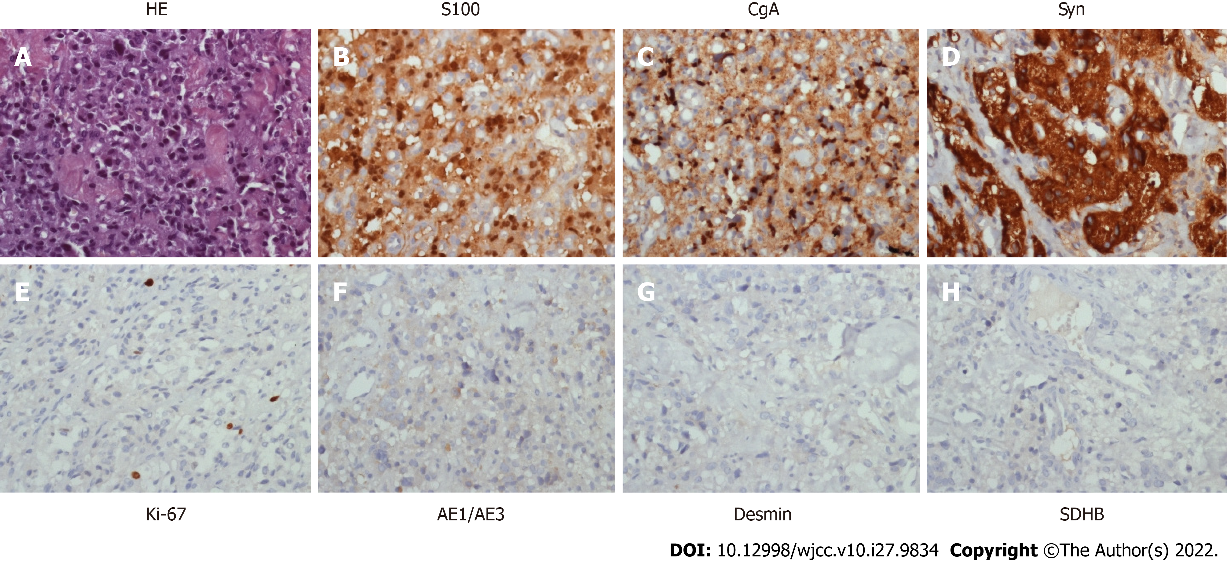
Figure 2 Representative immunohistochemical staining of the metastatic tumor.
A: HE-stained section; B: Cancer cells show positive staining for S100 in the cytoplasm and nucleus; C: Positive staining for CgA in the cytoplasm; D: Positive staining for Syn in the cytoplasm; E: Positive nuclear staining for Ki-67; F-H: Negative staining for AE1/AE3, Desmin, and SDHB in the cytoplasm of cancer cells (original magnification × 400).
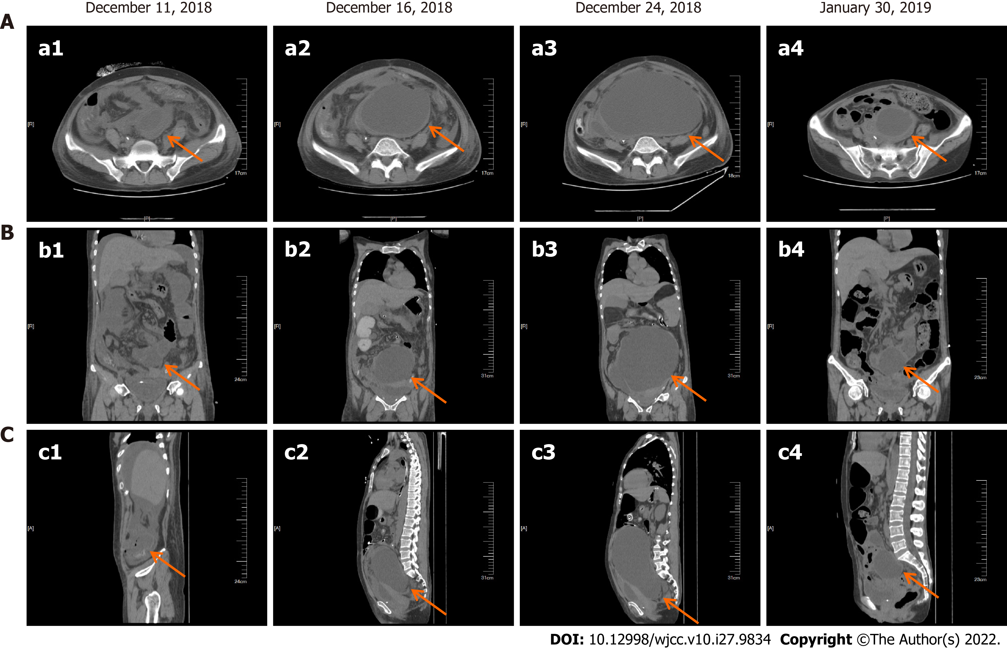
Figure 3 Representative radiologic images of the patient before and after chemotherapy.
A: Transverse plane; B: Coronal plane; C: Sagittal plane. a1-c1: December 11, 2018; a2-c2: December 16, 2018; a3-c3: December 24, 2018, before the treatment; a4-c4: January 30, 2019, after two cycles chemotherapy.
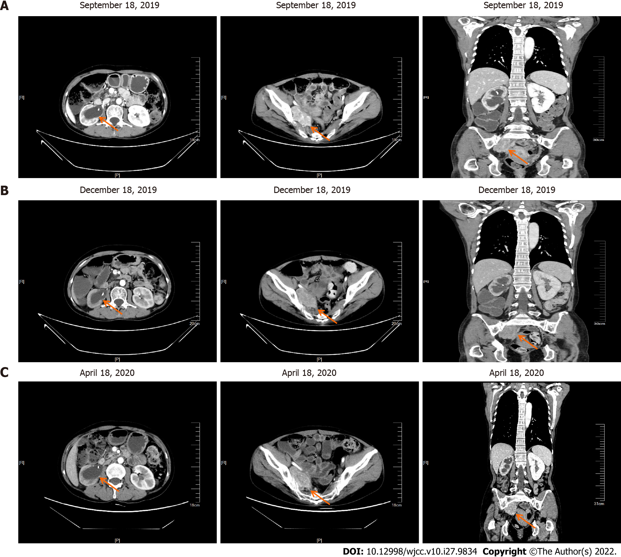
Figure 4 Follow-up results.
Enhanced computed tomography showing the same condition in the hydronephrosis of the right pelvis and renal calyces, accompanied with pelvic neoplasm. A: September 18, 2019; B: December 18, 2019; C: April 18, 2020.
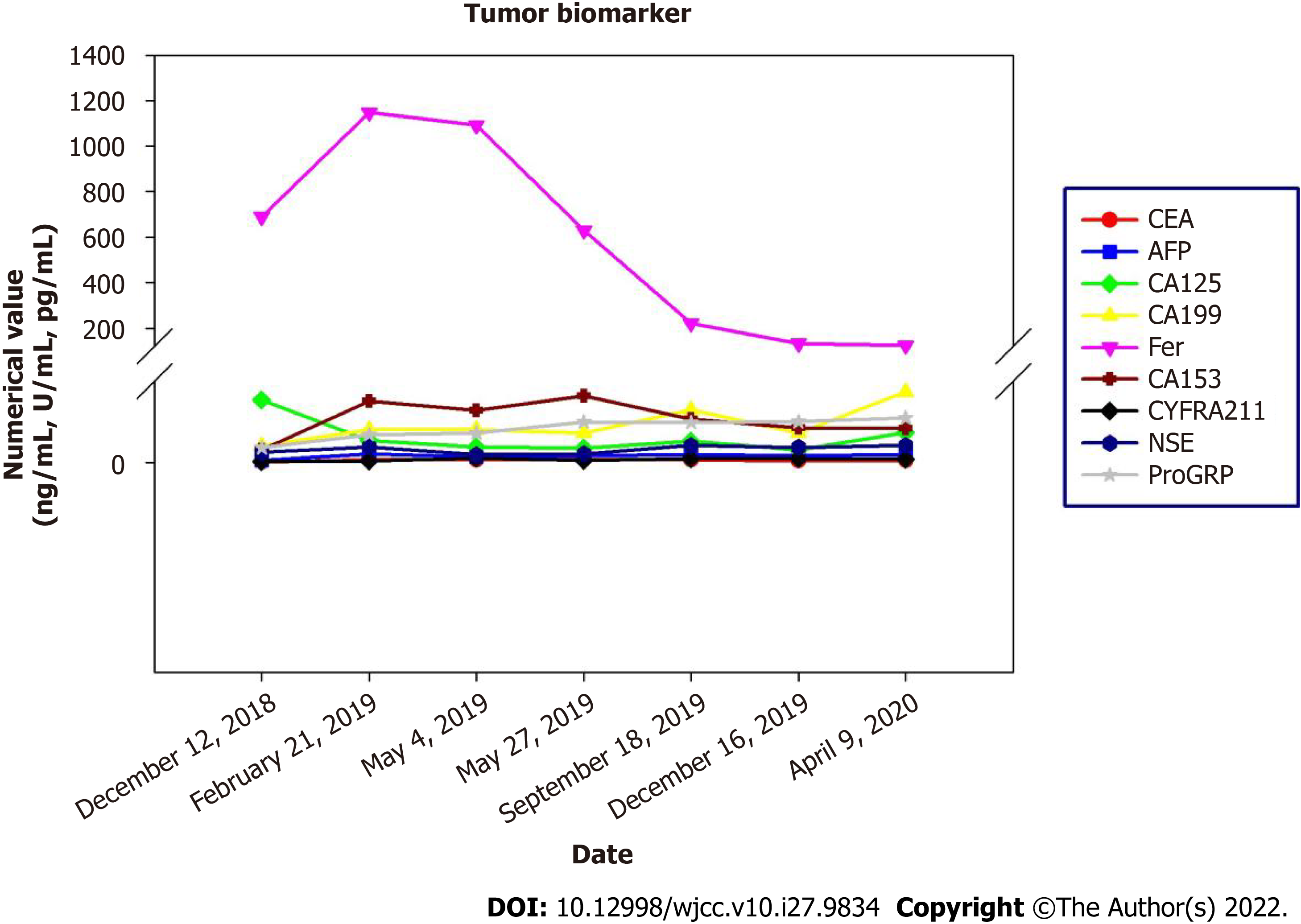
Figure 5 Blood tumor biomarkers.
CEA: Carcinoembryonic antigen; AFP: Acute flaccid paralysis; NSE: Neuron specific enolase.
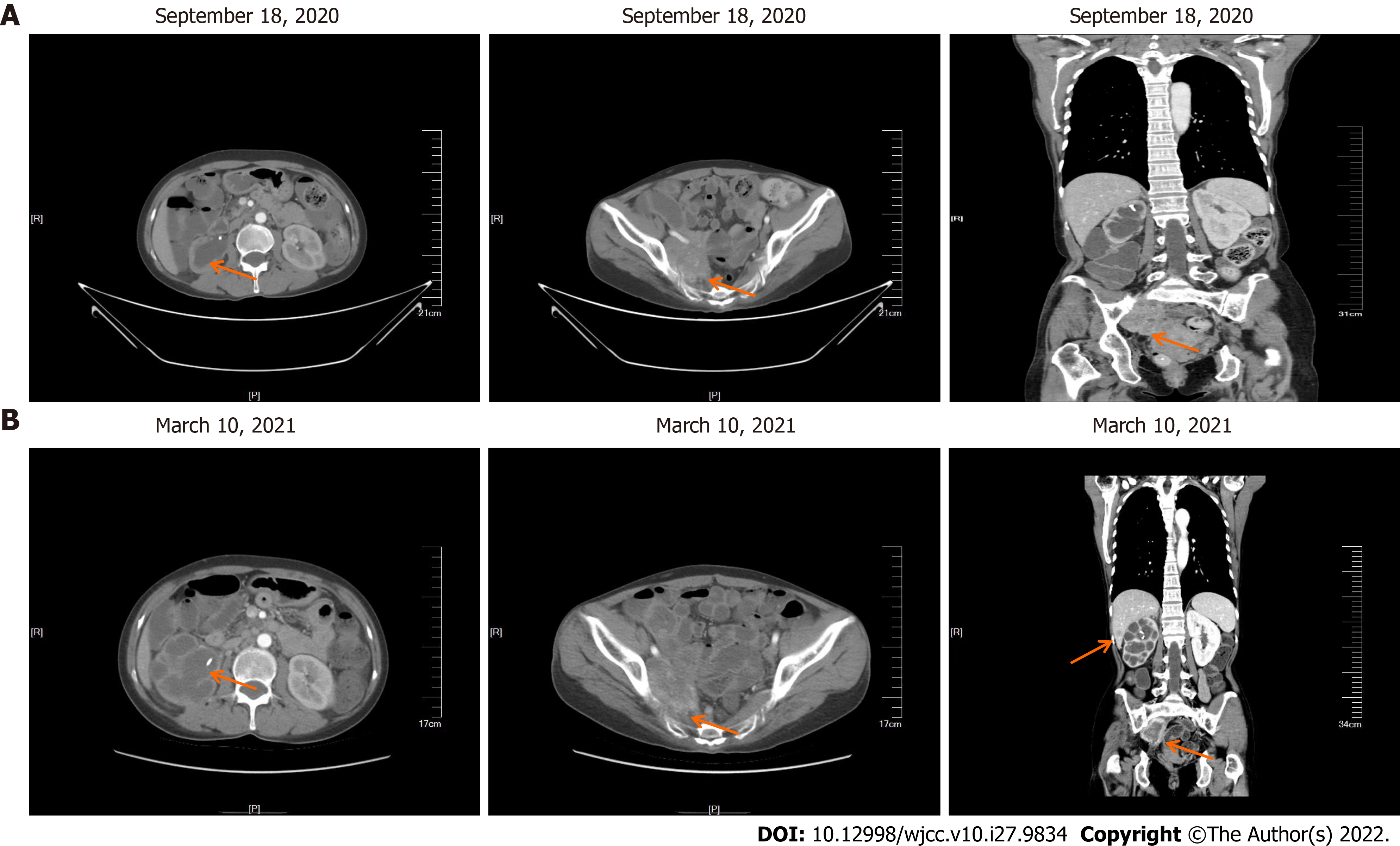
Figure 6 Follow-up imaging findings.
A: Enhanced computed tomography showing hydronephrosis of the right pelvis and renal calyces, accompanied with a larger pelvic neoplasm; B: Enhanced computed tomography scan showing multiple metastases in the liver.
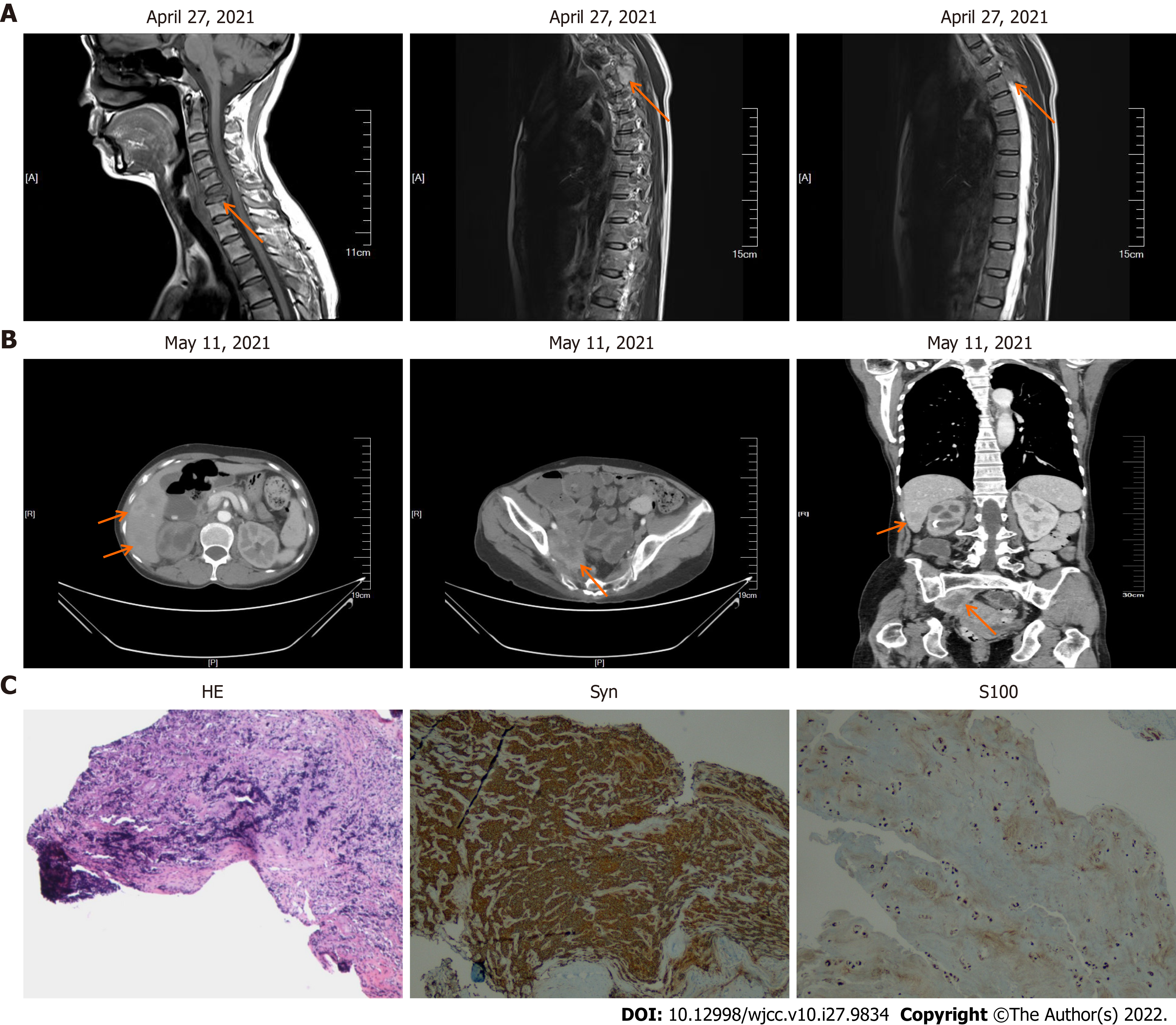
Figure 7 Follow-up imaging findings and representative immunohistochemical staining of the metastatic jugular tumor.
A: Enhanced computed tomography (CT) showing hydronephrosis of the right pelvis and renal calyces, accompanied with larger pelvic neoplasm; B: Enhanced CT scan showing multiple metastases in the liver; and C: Representative immunohistochemical staining of the metastatic jugular tumor: HE, cancer cells show positive staining for Syn in the cytoplasm, and positive staining for S100 in the cytoplasm and nucleus (magnification × 100).
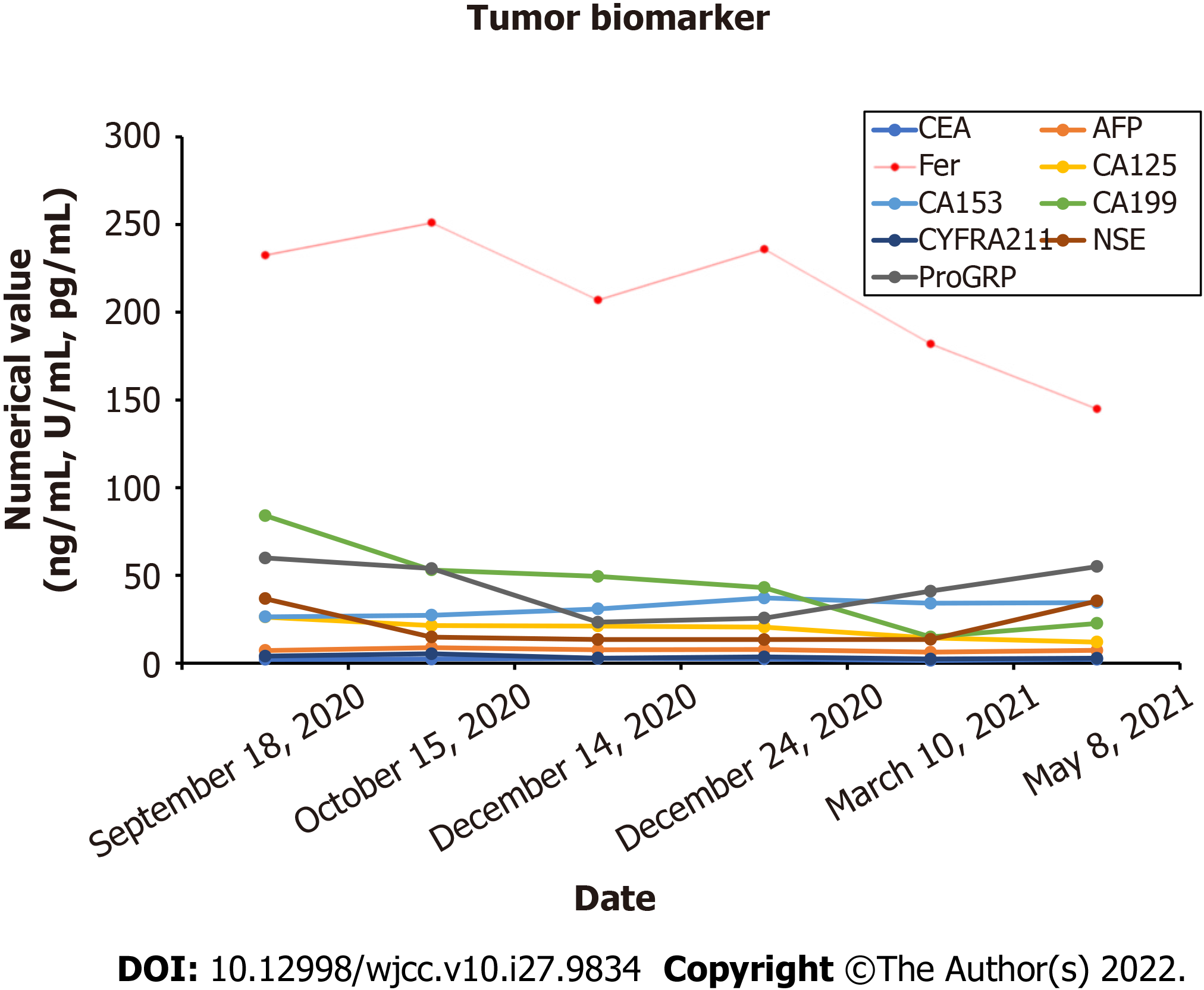
Figure 8 Blood tumor biomarkers after disease progression.
CEA: Carcinoembryonic antigen; AFP: Acute flaccid paralysis; NSE: Neuron specific enolase.
- Citation: Gan L, Shen XD, Ren Y, Cui HX, Zhuang ZX. Diagnostic features and therapeutic strategies for malignant paraganglioma in a patient: A case report. World J Clin Cases 2022; 10(27): 9834-9844
- URL: https://www.wjgnet.com/2307-8960/full/v10/i27/9834.htm
- DOI: https://dx.doi.org/10.12998/wjcc.v10.i27.9834


