Copyright
©The Author(s) 2022.
World J Clin Cases. Sep 26, 2022; 10(27): 9814-9820
Published online Sep 26, 2022. doi: 10.12998/wjcc.v10.i27.9814
Published online Sep 26, 2022. doi: 10.12998/wjcc.v10.i27.9814
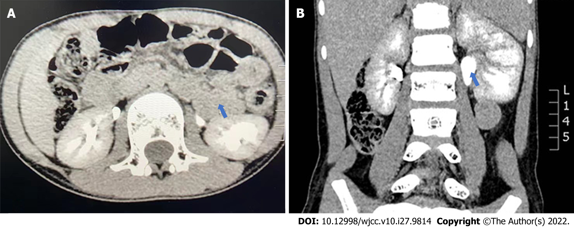
Figure 1 Contrast-enhanced computed tomography.
A: Computed tomography section at the level of renal vessels shows a horseshoe appearance of the bowel loops that contain the jejunal vessels radiating inside the hernial sac; B: Left hydronephrosis.
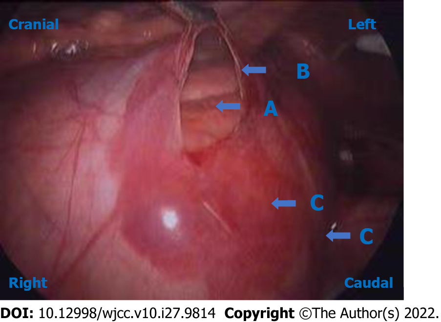
Figure 2 Intraoperative image.
The patient was lying on the right side while we performed the surgery. The observation hole was near the umbilicus. Intra-operatively, it was observed that small-bowel loops were lying within a hernial sac. The upper end of the ureter was located in the retroperitoneum, behind the hernia sac. The sac was opened with an ultrasonic shear, and the bowel was freed up. The hernia sac compressed the upper ureter of the left ureter and induced hydronephrosis. A: Small intestine; B: Hernia sac; C: Lateral peritoneum.
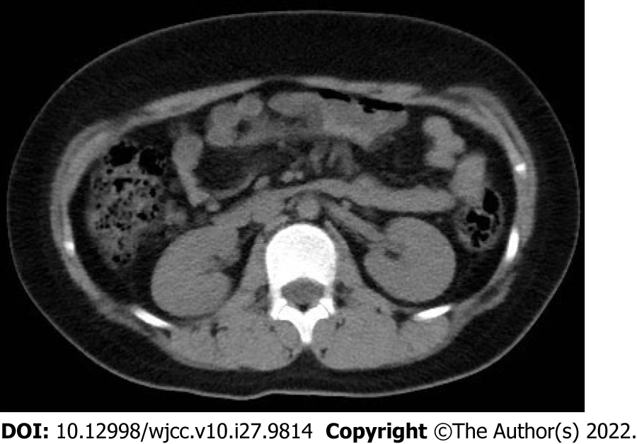
Figure 3 Computed tomography performed 2 years after laparoscopic paraduodenal hernia repair showed that the hydronephrosis was remitted and the paraduodenal hernia had been recovered.
- Citation: Wang X, Wu Y, Guan Y. Laparoscopic correction of hydronephrosis caused by left paraduodenal hernia in a child with cryptorchism: A case report. World J Clin Cases 2022; 10(27): 9814-9820
- URL: https://www.wjgnet.com/2307-8960/full/v10/i27/9814.htm
- DOI: https://dx.doi.org/10.12998/wjcc.v10.i27.9814


