Copyright
©The Author(s) 2022.
World J Clin Cases. Sep 26, 2022; 10(27): 9783-9789
Published online Sep 26, 2022. doi: 10.12998/wjcc.v10.i27.9783
Published online Sep 26, 2022. doi: 10.12998/wjcc.v10.i27.9783
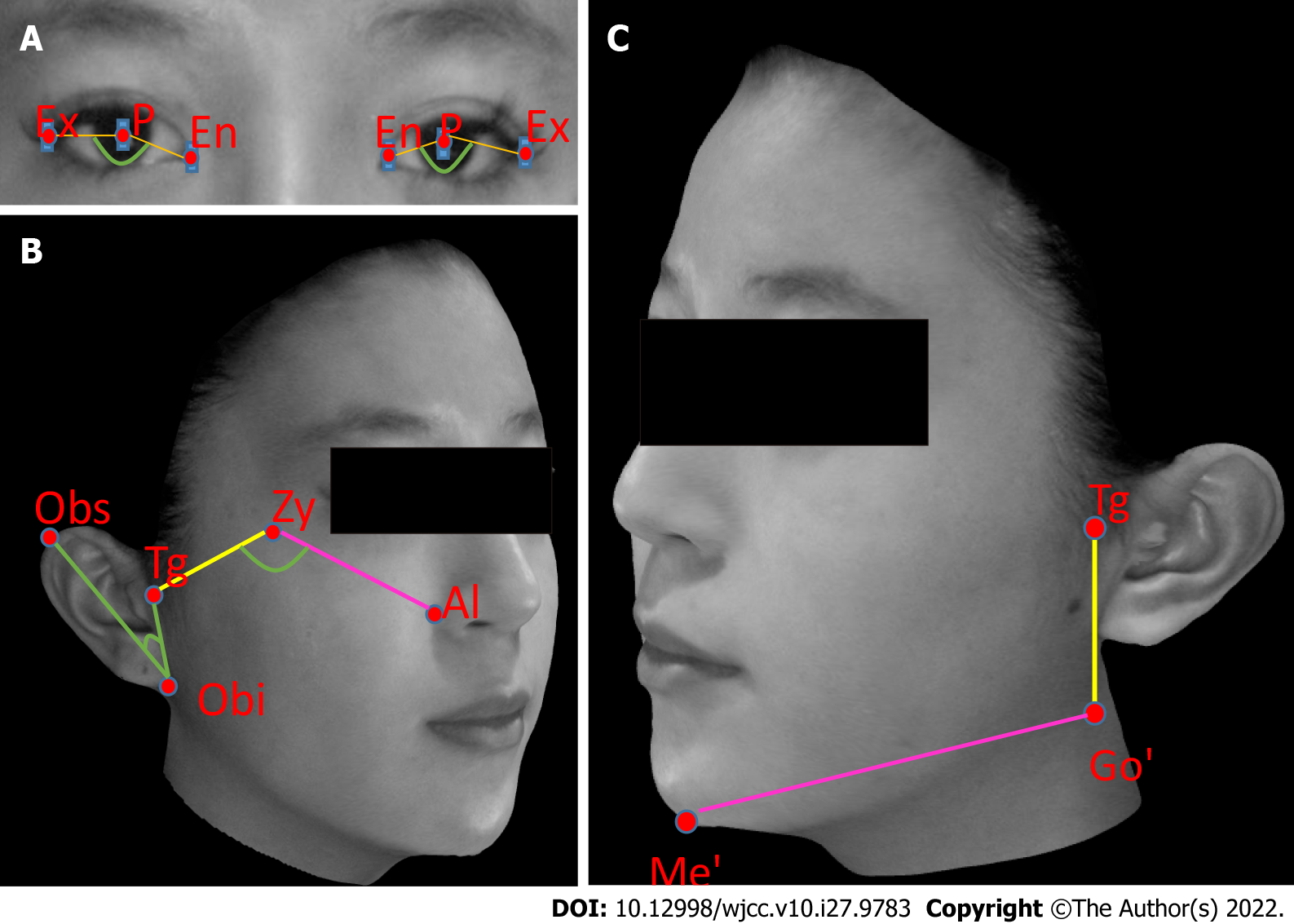
Figure 1 Illustrations of Morpheus three-dimensional camera measurements, showing facial landmarks used for Morpheus 3D Superimposition.
A: Angle formed by exocanthion-pupil-endocanthion for improvement in the orbital region; B: Angle formed by tragion-zygion-ala for improvement in the zygomatic region (right), using the angle formed by otobasion superius-otobasion inferius-tragion as reference; C: Distance between soft tissue gonion and soft tissue menton for improvements in the mandibular region (left), using the distance between tragion and soft tissue gonion as reference. Al: Ala; En: Endocanthion; Ex: Exocanthion; Go’: Soft tissue gonion; Me’: Soft tissue menton; Obi: Otobasion inferius; Obs: Otobasion superius; P: Pupil; Tg: Tragion; Zy: Zygion.
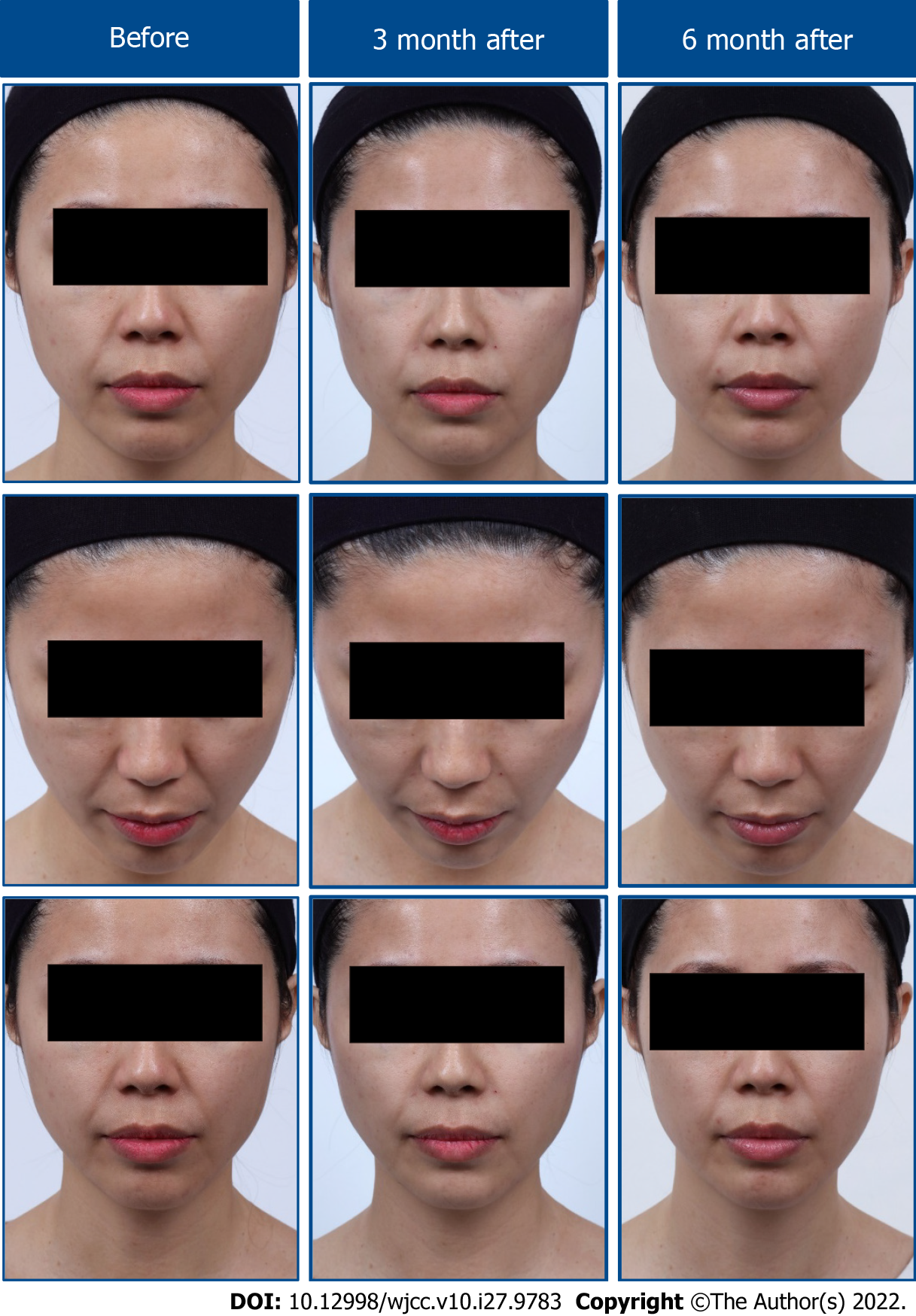
Figure 2 Representative photographs showing the effects of nonsurgical facial retightening procedure (True Lift®) with high G’ filler (Restylane® Lyft Lidocaine) on overall changes of the facial firmness at 3-mo and 6-mo post-injection.
Case patient: 29-years-old.
- Citation: Huang P, Li CW, Yan YQ. Efficacy evaluation of True Lift®, a nonsurgical facial ligament retightening injection technique: Two case reports. World J Clin Cases 2022; 10(27): 9783-9789
- URL: https://www.wjgnet.com/2307-8960/full/v10/i27/9783.htm
- DOI: https://dx.doi.org/10.12998/wjcc.v10.i27.9783


