Copyright
©The Author(s) 2022.
World J Clin Cases. Sep 26, 2022; 10(27): 9760-9767
Published online Sep 26, 2022. doi: 10.12998/wjcc.v10.i27.9760
Published online Sep 26, 2022. doi: 10.12998/wjcc.v10.i27.9760
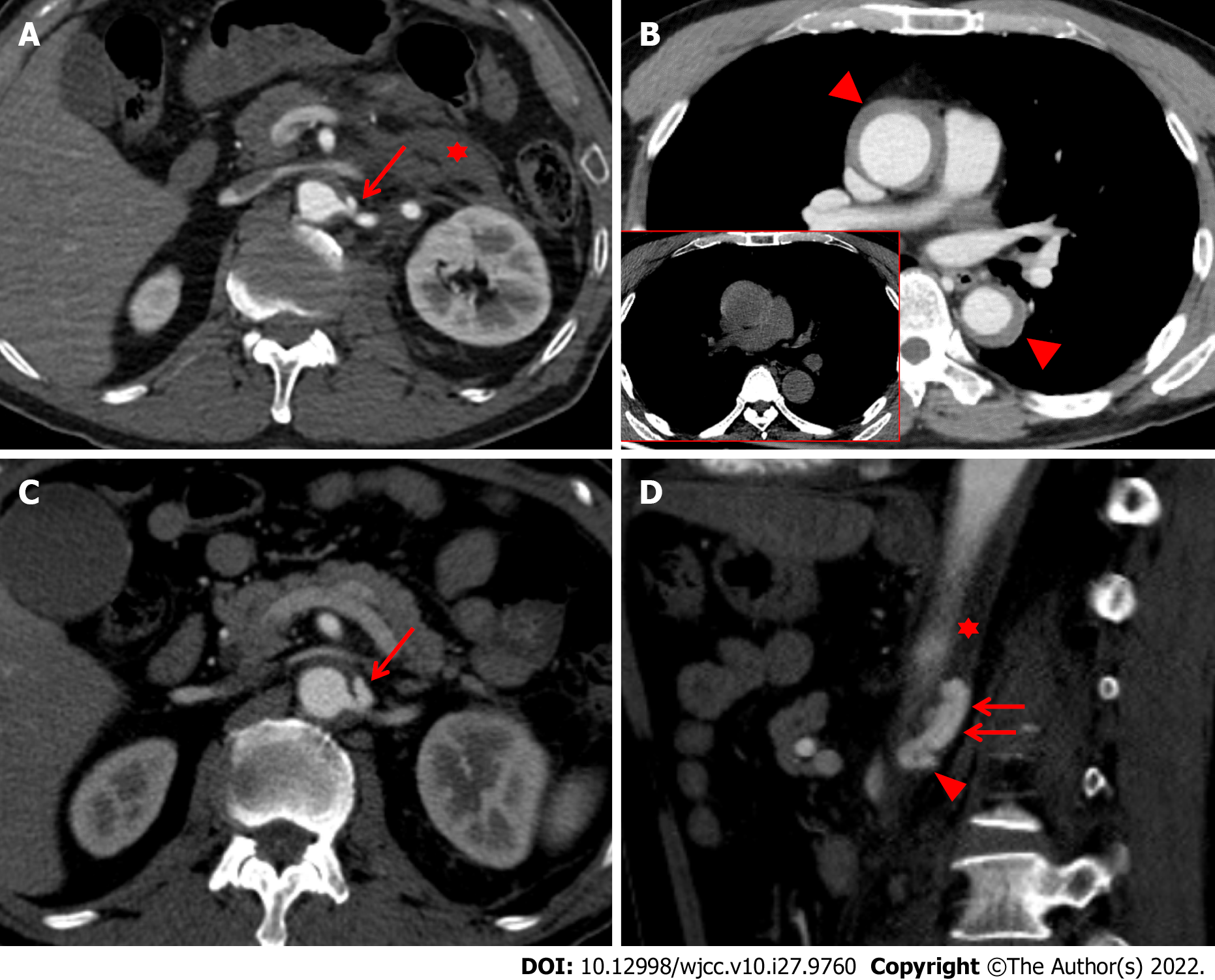
Figure 1 Initial computed tomography scans taken 1 h (A and B) after injury, and follow-up CT scans taken 15 d (C and D) after injury.
A: The initial abdominal computed tomography (CT) scan also revealed focal out-pouching enhancing nodular lesion, suggesting a pseudoaneurysm (red arrow) arising from the proximal portion of the left renal artery with retroperitoneal hematoma (red asterisk); B: The initial chest CT scan taken one hour after injury showed Stanford type A intramural hematoma (IMH) (red arrowheads), which represent slightly hyperdensity on precontrast image (figure in red-border box); C: The CT scan reveals an increase in the size and extent of the pseudoaneurysm (red arrow) on the axial image; D: The pseudoaneurysm arising from the proximal renal artery (red arrowhead) was localized within aortic media (red asterisk), resulting in an intramural blood collection (red arrows) seen on the sagittal image.
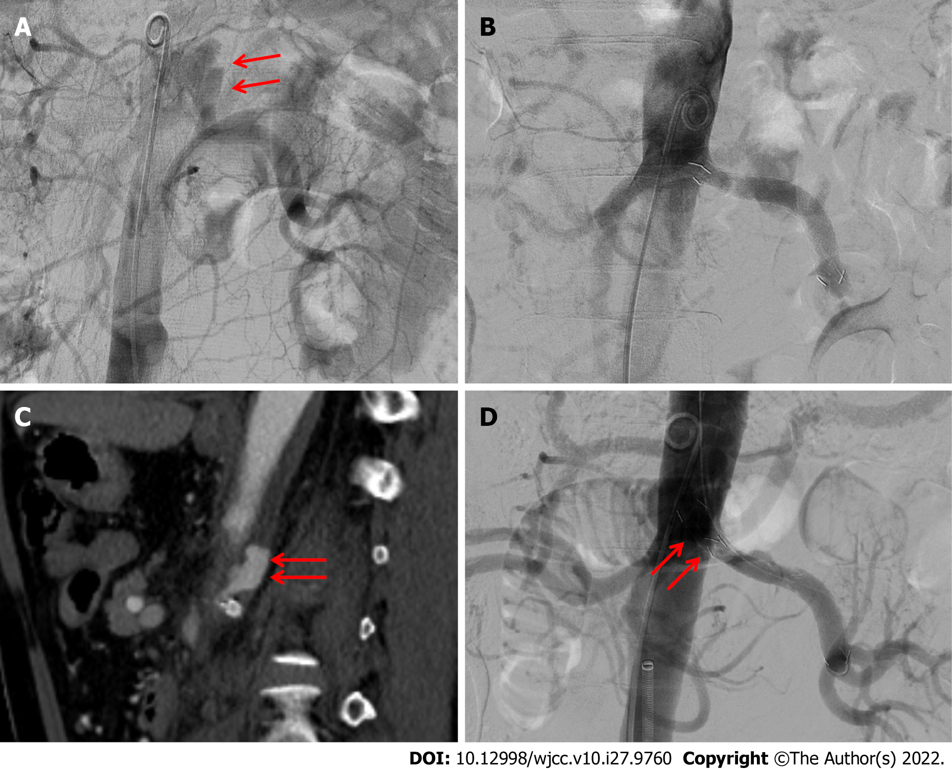
Figure 2 Endovascular procedure performed 15 d and 17 d after injury.
A: The pseudoaneurysm (red arrow) arising from the left proximal renal artery was detected using abdominal aortography 15 d after injury; B: An 8 mm × 55 mm covered stent was deployed along the left renal main artery, bridging the pseudoaneurysm and covering the parent artery, successfully excluding the pseudoaneurysm; exclusion was confirmed using final aortography; C: Follow-up CT scan two days after the first procedure reveals a decrease in the size of the pseudoaneurysm (red arrow) arising from the renal artery; D: Second endovascular procedure performed three days after the first treatment. The additional covered stent (red arrows) was placed to fully cover the orifice of the left renal artery to prevent 1A endoleak, successfully excluding the pseudoaneurysm, as confirmed using final aortography.
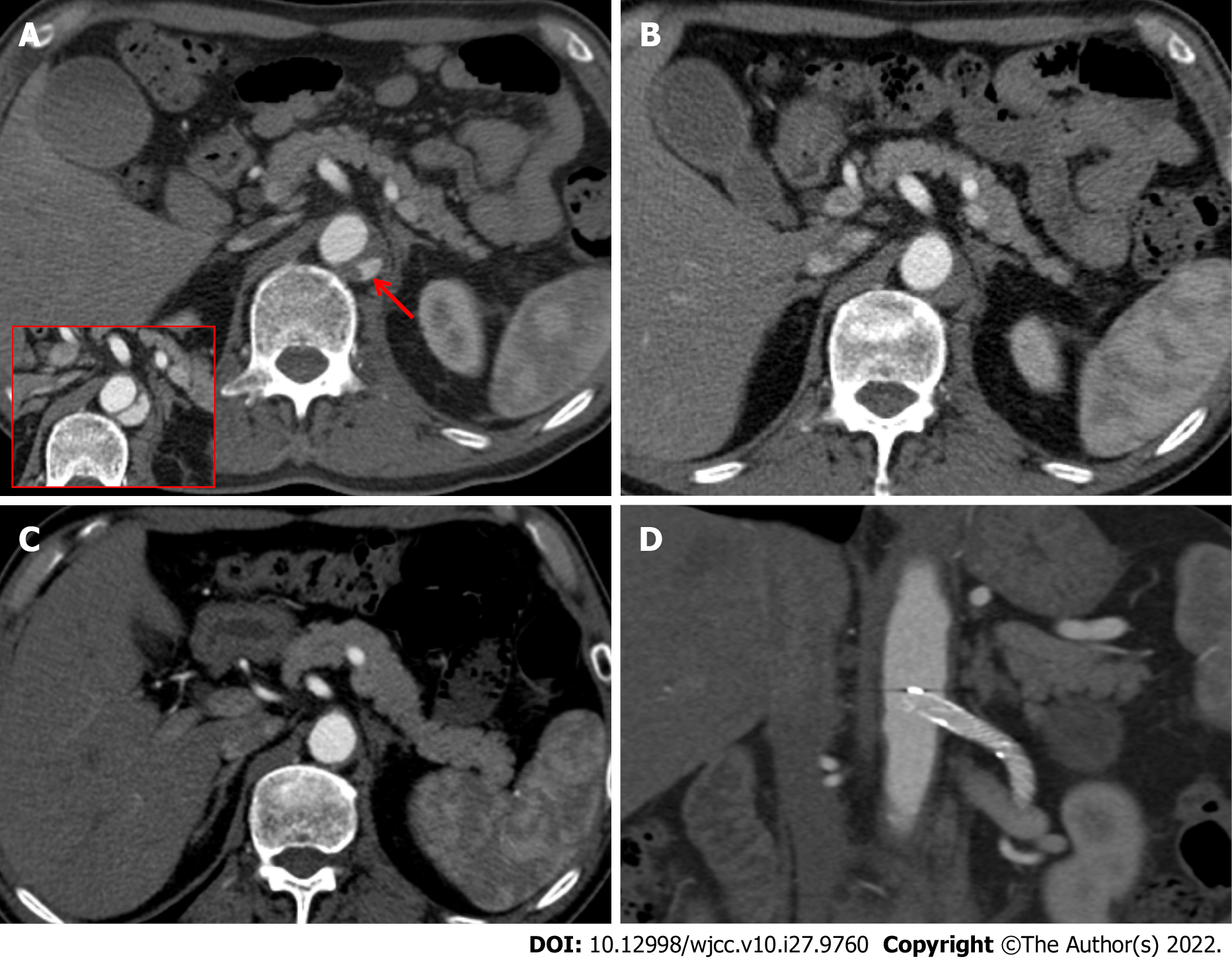
Figure 3 Consecutive computed tomography scans performed 26 d, 45 d, and 7 mo after injury.
A: The computed tomography (CT) scan taken 26 d after injury revealed a decrease in the size and extent of the intramural blood collection (red arrow) compared with the CT scan taken seven days prior (figure in red-border box); B: The CT scan taken 45 d after injury showed that the intramural blood collection markedly decreased in size; C: In the 7-mo CT scan, the intramural hematoma (IMH) had completely disappeared; D: There was no evidence of stent-related complications such as perforation, obstruction, and stent migration.
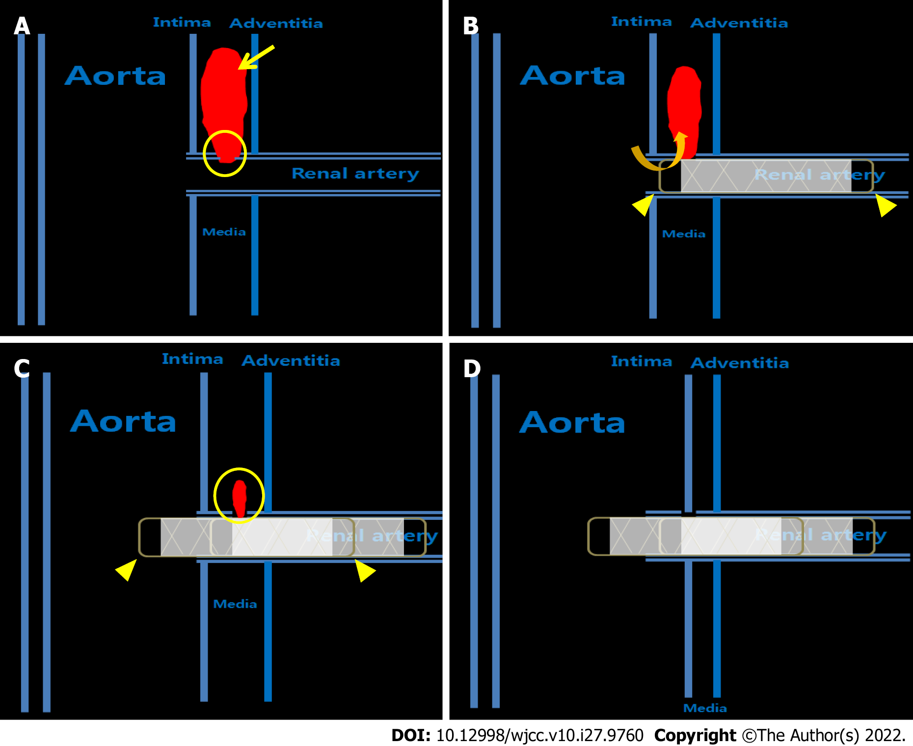
Figure 4 Two-dimensional illustration of the treatment process for aortic intramural hematoma associated with a pseudoaneurysm arising from the renal artery after blunt trauma using covered stents.
A: The intimomedial tear (yellow circle) of the proximal renal artery occurs first by trauma, leading to intramural blood collection, and finally within the intramural hematoma (yellow asterisk). This intramural blood collection is defined as a pseudoaneurysm (yellow arrow); B: Although the size of the pseudoaneurysm appeared to decrease after the deployment of the stent graft, a large blood pool was still present due to the endoleak caused by the residual blood flow (orange angled arrow) through the space between the uncovered area of the stent graft (yellow arrowhead) and intimomedial flap; C: The intramural blood collection (yellow circle) was markedly decreased in size and extent after installing an additional stent graft (yellow arrowhead); D: The intramural blood collection and hematoma completely disappeared seven months after the second session.
- Citation: Kim Y, Lee JY, Lee JS, Ye JB, Kim SH, Sul YH, Yoon SY, Choi JH, Choi H. Endovascular treatment of traumatic renal artery pseudoaneurysm with a Stanford type A intramural haematoma: A case report. World J Clin Cases 2022; 10(27): 9760-9767
- URL: https://www.wjgnet.com/2307-8960/full/v10/i27/9760.htm
- DOI: https://dx.doi.org/10.12998/wjcc.v10.i27.9760


