Copyright
©The Author(s) 2022.
World J Clin Cases. Sep 26, 2022; 10(27): 9734-9742
Published online Sep 26, 2022. doi: 10.12998/wjcc.v10.i27.9734
Published online Sep 26, 2022. doi: 10.12998/wjcc.v10.i27.9734
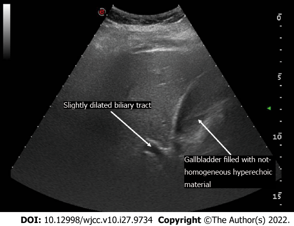
Figure 1 Ultrasound scan.
Distended gallbladder filled with non-homogeneous hyperechoic material and slightly dilated intrahepatic biliary tract, the common bile duct was not visible due to intestinal gas.
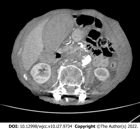
Figure 2 Computed tomography scan of intra- and extra-hepatic biliary ducts demonstrated wider dilatation.
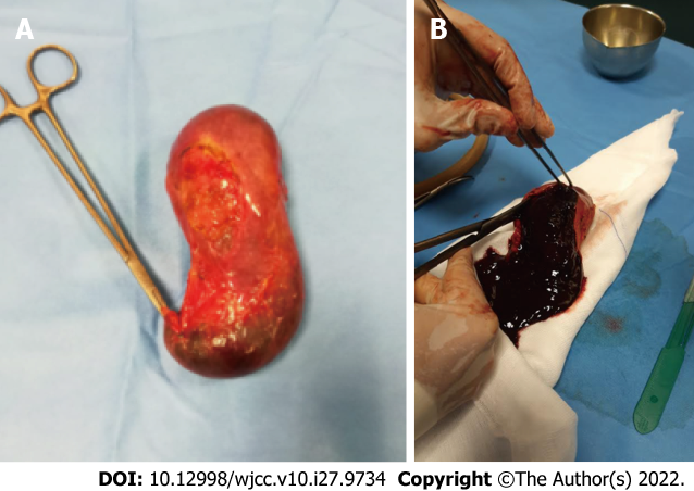
Figure 3 Surgical specimen.
A: When open cholecystectomy was performed, choledocotomy with Kehr tube apposition completed the surgery due to dilated hepatocoledocus (approximately 25 mm); B: When the gallbladder was inspected at the backtable, it appeared entirely occupied by clots.
- Citation: Valenti MR, Cavallaro A, Di Vita M, Zanghi A, Longo Trischitta G, Cappellani A. Gallbladder hemorrhage–An uncommon surgical emergency: A case report. World J Clin Cases 2022; 10(27): 9734-9742
- URL: https://www.wjgnet.com/2307-8960/full/v10/i27/9734.htm
- DOI: https://dx.doi.org/10.12998/wjcc.v10.i27.9734


