Copyright
©The Author(s) 2022.
World J Clin Cases. Sep 26, 2022; 10(27): 9628-9640
Published online Sep 26, 2022. doi: 10.12998/wjcc.v10.i27.9628
Published online Sep 26, 2022. doi: 10.12998/wjcc.v10.i27.9628
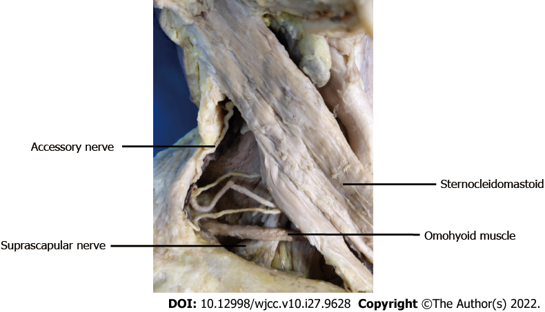
Figure 1 The suprascapular nerve beneath the lower belly of the omohyoid muscle.
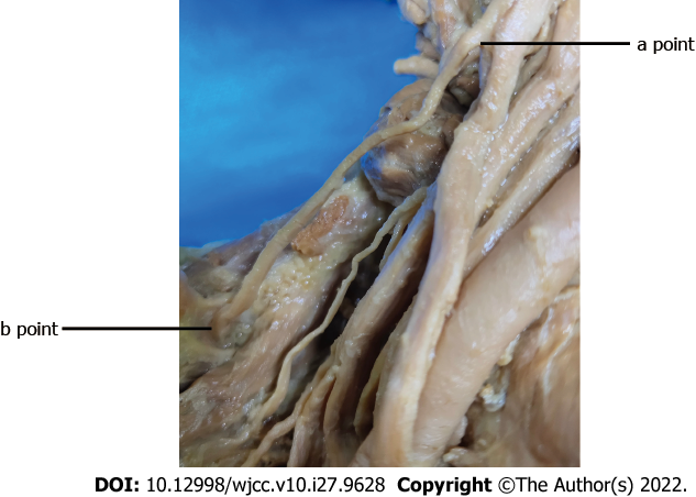
Figure 2 Suprascapular nerve from the origin of the brachial plexus (a point) to the scapular notch (b point).
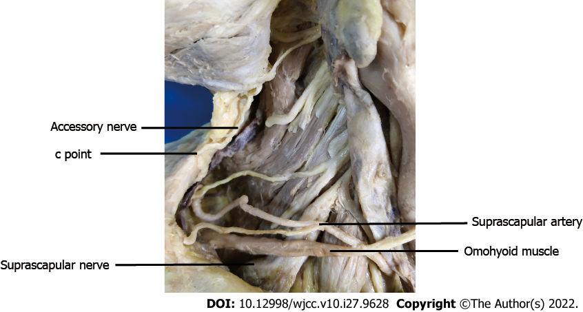
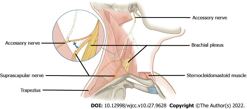
Figure 3 Relationship between the suprascapular nerve and the accessory nerve.
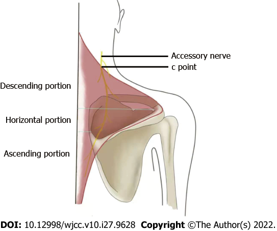
c point was referring to the position where accessory nerve entered into the border of the trapezius muscle.
Figure 4 Schematic diagram of distribution of accessory nerve in trapezius muscle.
Figure 5 Schematic diagram of anastomosis between the proximal stump of the accessory nerve and the suprascapular nerve.
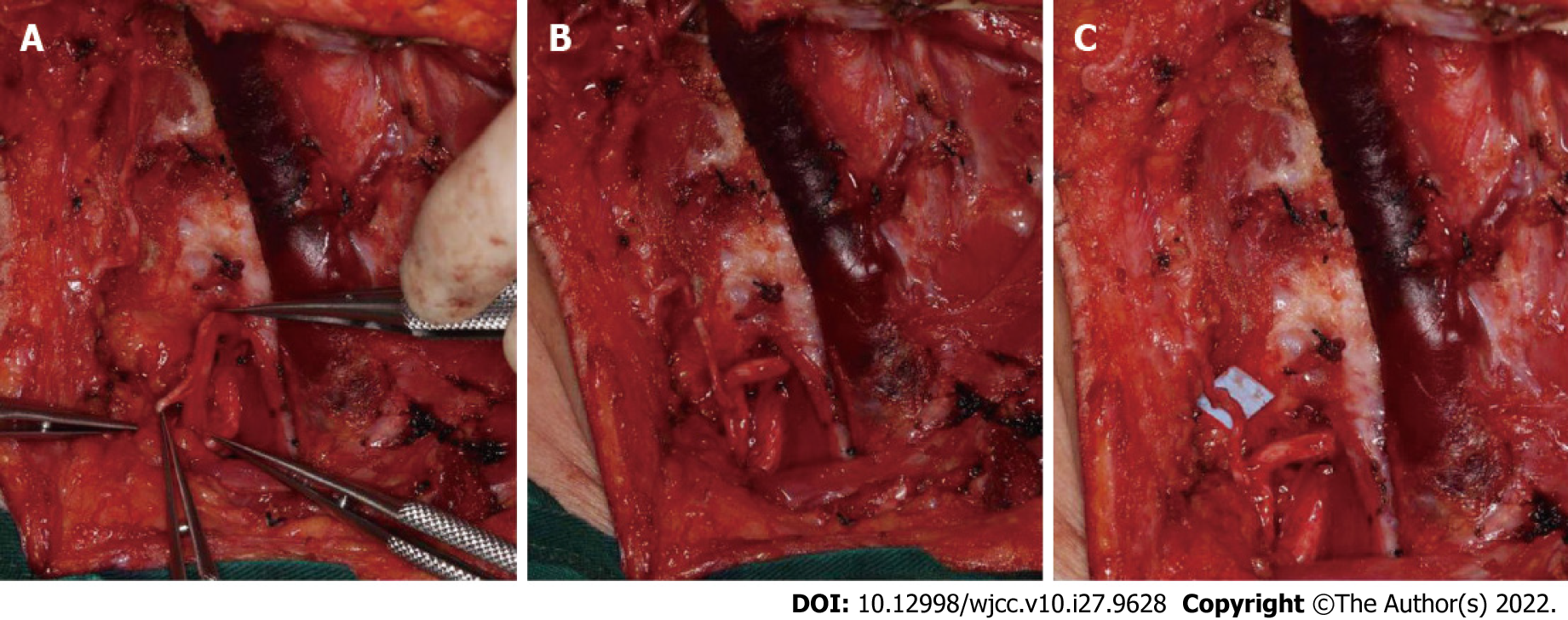
Figure 6 Transfer procedure between the suprascapular nerve and the proximal stump of accessory nerve.
A: One third of the diameter of the suprascapular nerve (SCN) was dissected; B: The nerve was cut off and put next to the proximal stump of accessory nerve (AN); C: The two stumps of AN and SCN were sutured with nylon suture (8-0).
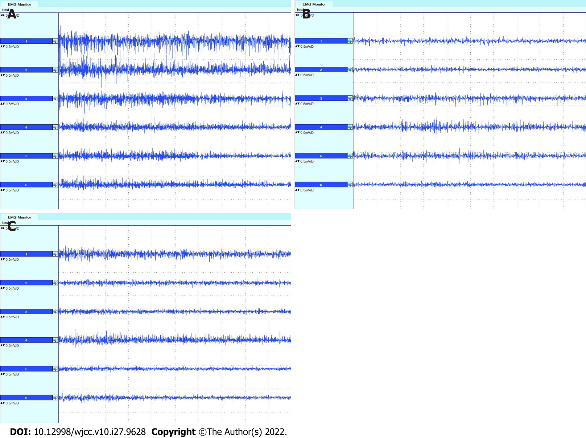
Figure 7 Electromyographs of the trapezius muscle.
A: Before surgery; B: Three months after surgery; C: Nine months after surgery. Electromyographs from top to bottom show the results of the descending portion on the left side, the descending portion on the right side, the horizontal portion on the left side, the horizontal portion on the right side, the ascending portion on the left side, and the horizontal portion on the right side, respectively.
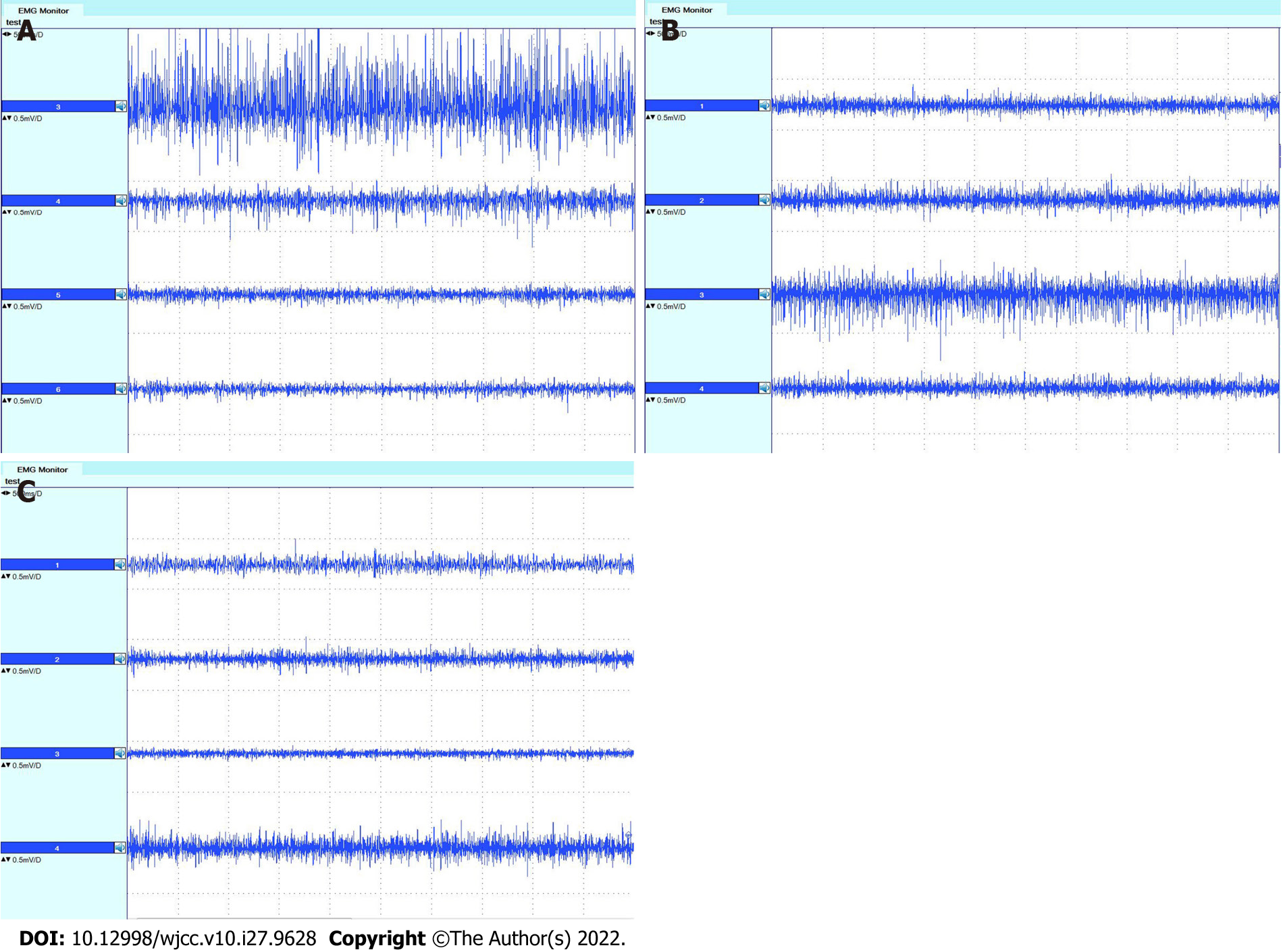
Figure 8 Electromyographs of the supraspinatus and infraspinatus muscles.
A: Before surgery; B: Three months after surgery; C: Nine months after surgery. Electromyographs from top to bottom show the results of the supraspinatus on the left side, supraspinatus on the right side, infraspinatus on the left side, and infraspinatus on the right side, respectively.
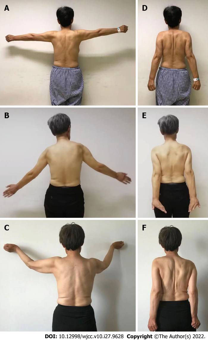
Figure 9 The patient’s clinical manifestation of upper limb movement.
A and D: Before surgery; B and E: Three months after surgery; C and F: Nine months after surgery. The panels A-C showed the movement of abduction of the upper arms, and the panels D-F showed the movement of external rotation of the upper arms.
- Citation: Wang JW, Zhang WB, Li F, Fang X, Yi ZQ, Xu XL, Peng X, Zhang WG. Anatomy and clinical application of suprascapular nerve to accessory nerve transfer. World J Clin Cases 2022; 10(27): 9628-9640
- URL: https://www.wjgnet.com/2307-8960/full/v10/i27/9628.htm
- DOI: https://dx.doi.org/10.12998/wjcc.v10.i27.9628


