Copyright
©The Author(s) 2022.
World J Clin Cases. Sep 6, 2022; 10(25): 9087-9095
Published online Sep 6, 2022. doi: 10.12998/wjcc.v10.i25.9087
Published online Sep 6, 2022. doi: 10.12998/wjcc.v10.i25.9087
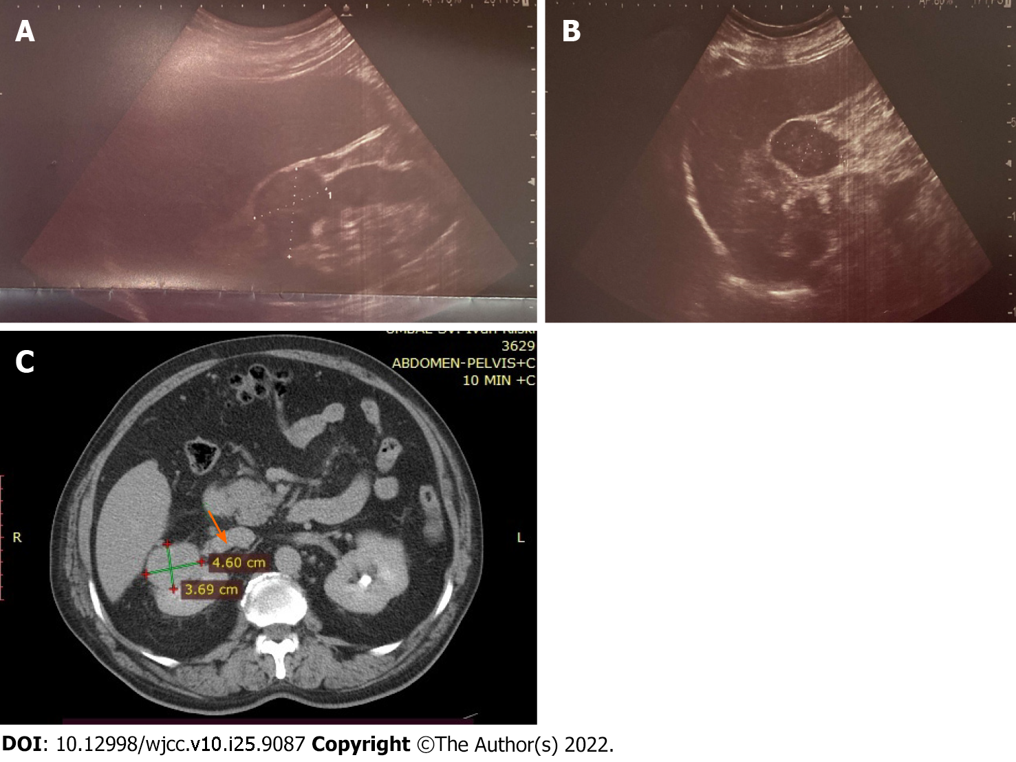
Figure 1 Imaging findings of the patient’s kidney tumor.
A and B: Representative abdominal ultrasound images showing the liver and right kidney with tumor formation; C: Image from the contrast-enhanced computed tomography of the patient showing right kidney with tumor formation and tumor involvement of the vena cava inferior (arrow).
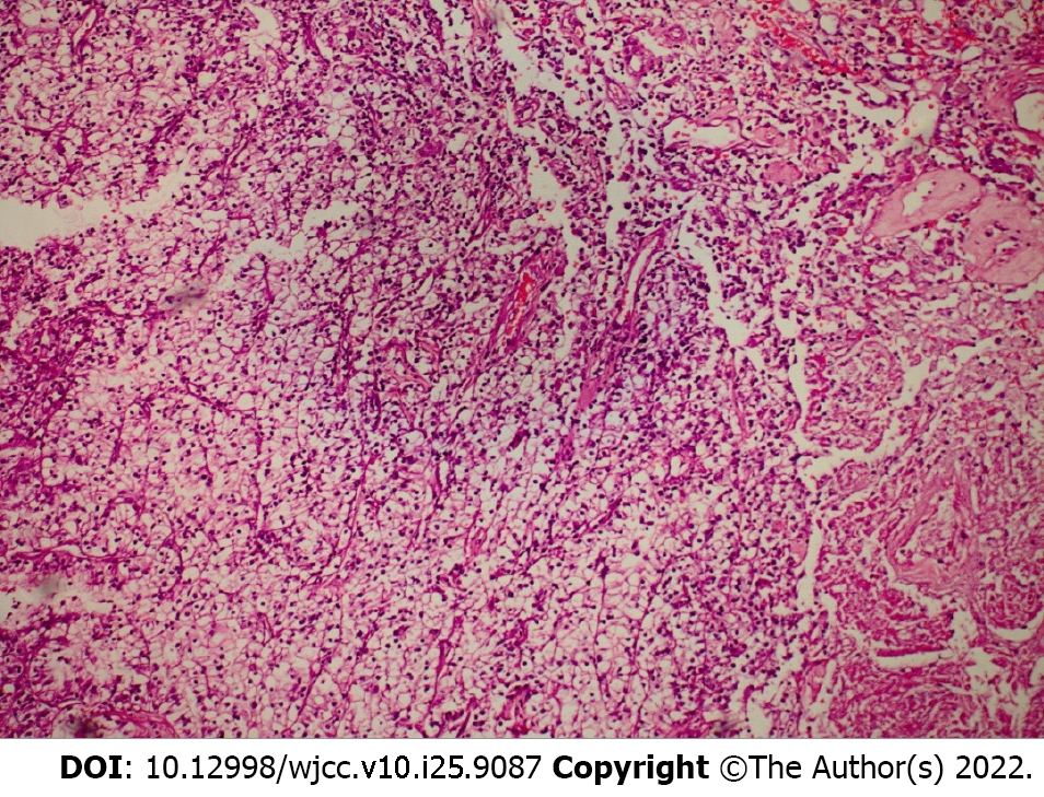
Figure 2 Histology findings of the patient’s kidney tumor.
Clear cell renal cell carcinoma was observed, of Grade 2 (World Health Organization/International Society of Urological Pathology) and with focal areas of necrosis. Hematoxylin and eosin stain, 100 × magnification.
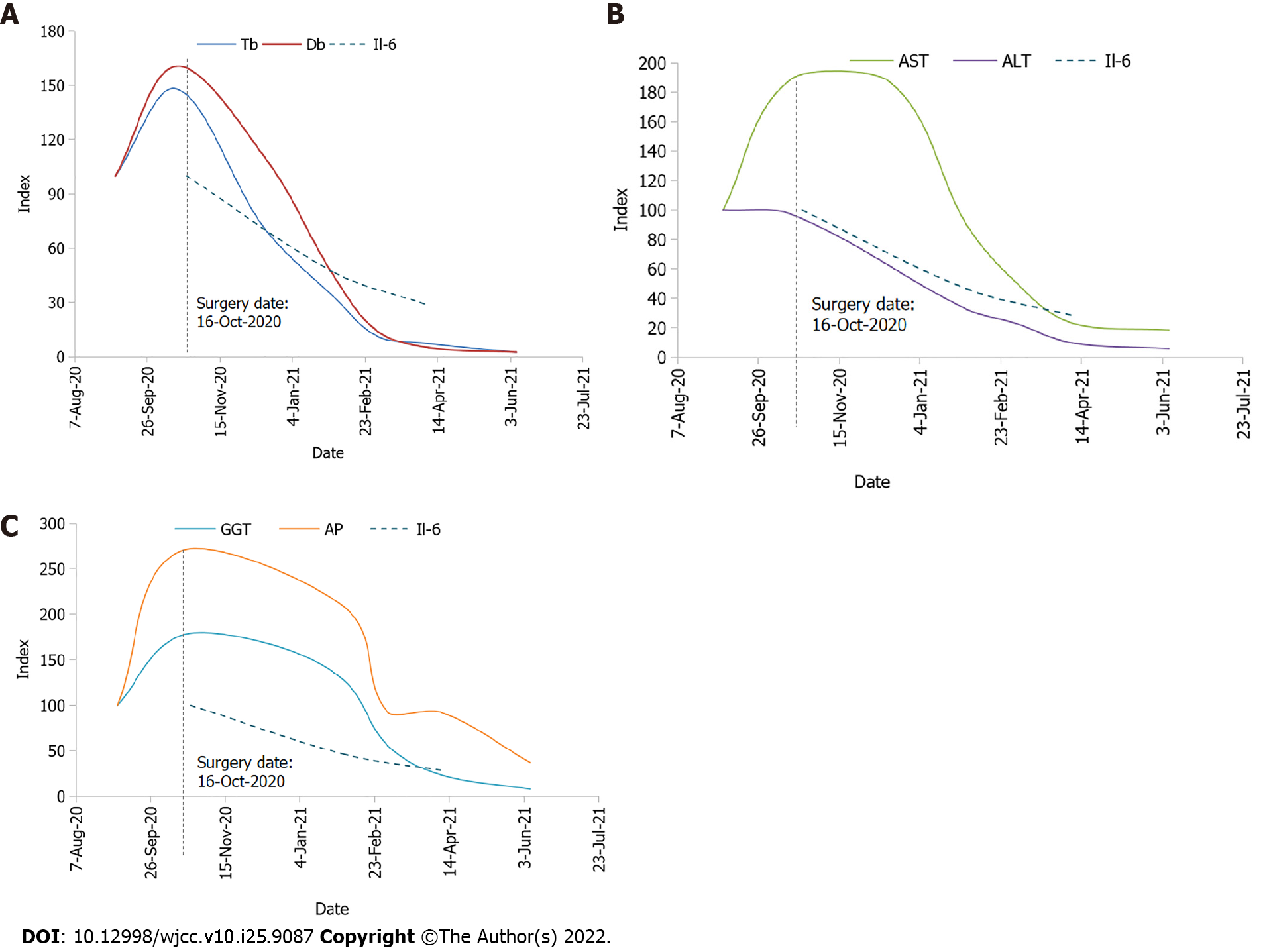
Figure 3 Dynamics of key laboratory parameters.
A: Total bilirubin (Tb), direct bilirubin (Db) and interleukin (IL)-6; B: Aspartate aminotransferase (AST), alanine aminotransferase (ALT) and IL-6; C: Gamma-glutamyl transferase (GGT), alkaline phosphatase (AP) and IL-6. Values were indexed to 100 as of the first available date for Tb, Db, AST, ALT, GGT, AP, and IL-6. The date of the surgery was October 16, 2020, with the following dates denoted by a dotted vertical line on the chart. The graphs only show values measured at key events.
- Citation: Popov DR, Antonov KA, Atanasova EG, Pentchev CP, Milatchkov LM, Petkova MD, Neykov KG, Nikolov RK. Renal cell carcinoma presented with a rare case of icteric Stauffer syndrome: A case report. World J Clin Cases 2022; 10(25): 9087-9095
- URL: https://www.wjgnet.com/2307-8960/full/v10/i25/9087.htm
- DOI: https://dx.doi.org/10.12998/wjcc.v10.i25.9087


