Copyright
©The Author(s) 2022.
World J Clin Cases. Aug 26, 2022; 10(24): 8788-8796
Published online Aug 26, 2022. doi: 10.12998/wjcc.v10.i24.8788
Published online Aug 26, 2022. doi: 10.12998/wjcc.v10.i24.8788
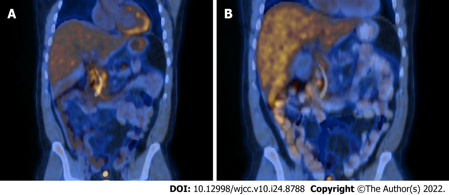
Figure 1 Fludeoxy glucose-positron emission tomography/computed tomography on diagnosis.
A: The image shows diffusely increased tracer activity within the pancreas suggestive of an inflammatory process rather than malignancy; B: Eight weeks of tapered prednisone.
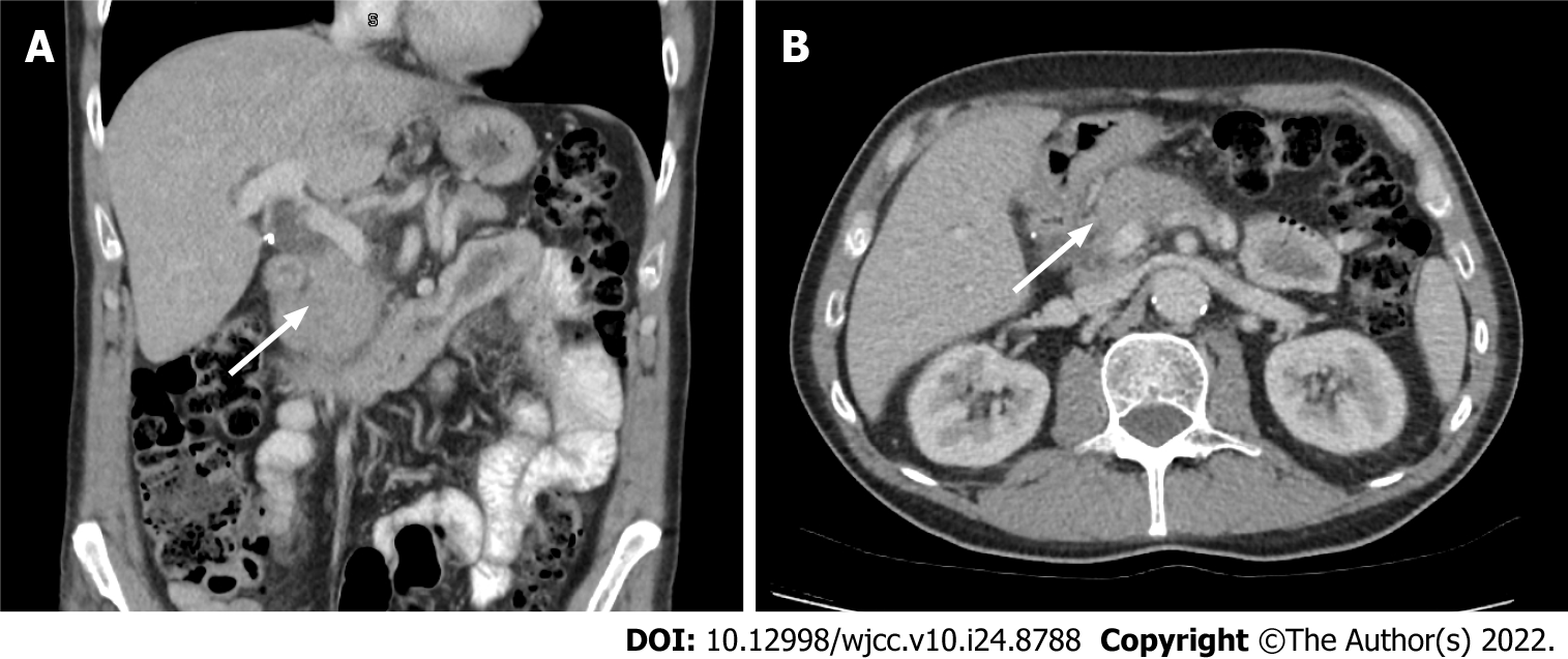
Figure 2 Computed tomography abdomen of case 2.
A: Mild thickening and oedema of the head and body of the pancreas with subtle effacement of the peri-pancreatic fat planes coronal section is demonstrated; B: Axial section.
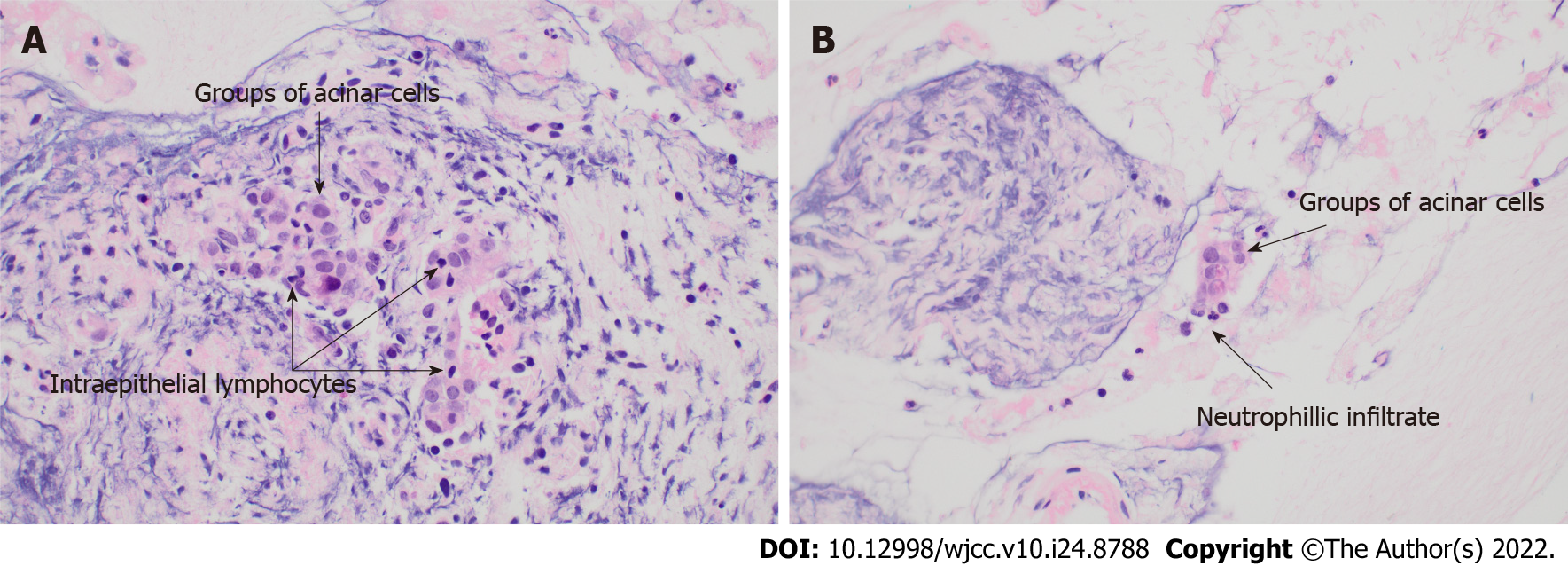
Figure 3 Histopathology of case 3.
A: Acinar cells with intraepithelial lymphocytes present, consistent with lymphocytic acinar inflammation; B: Neutrophils in the close vicinity of the acinar cell groups, suggestive of granulocytic acinar infiltrate.
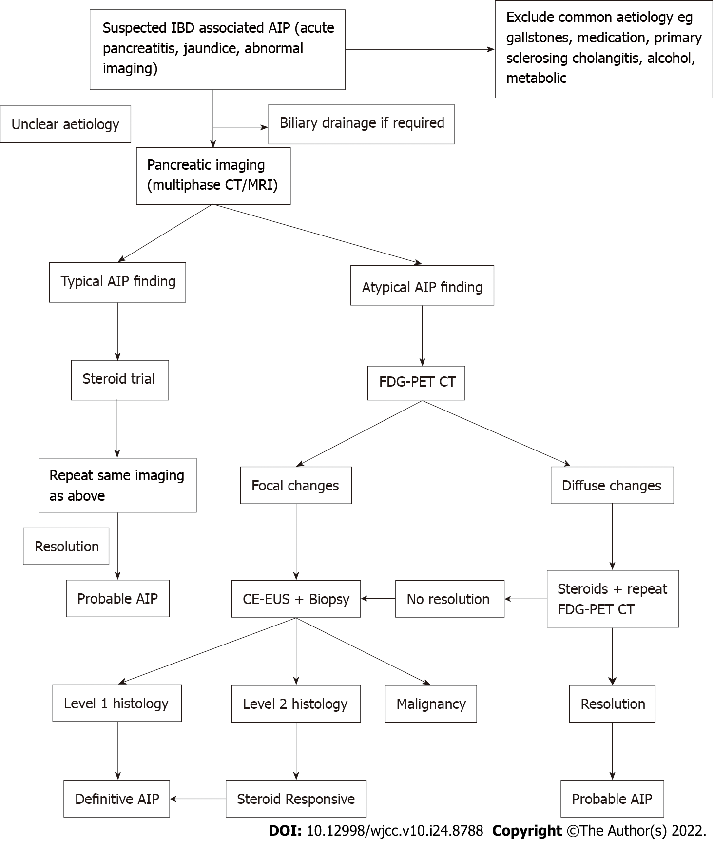
Figure 4 Suggested diagnostic algorithm for autoimmune pancreatitis in inflammatory bowel diseases.
IBD: Inflammatory bowel diseases; AIP: Autoimmune pancreatitis; CT: Computed tomography; MRI: Magnetic resonance imaging; FDG-PET: Fludeoxy glucose-positron emission tomography; CE-EUS: Contrast enhanced endoscopic ultrasound.
- Citation: Ghali M, Bensted K, Williams DB, Ghaly S. Type 2 autoimmune pancreatitis associated with severe ulcerative colitis: Three case reports. World J Clin Cases 2022; 10(24): 8788-8796
- URL: https://www.wjgnet.com/2307-8960/full/v10/i24/8788.htm
- DOI: https://dx.doi.org/10.12998/wjcc.v10.i24.8788


