Copyright
©The Author(s) 2022.
World J Clin Cases. Aug 26, 2022; 10(24): 8761-8767
Published online Aug 26, 2022. doi: 10.12998/wjcc.v10.i24.8761
Published online Aug 26, 2022. doi: 10.12998/wjcc.v10.i24.8761
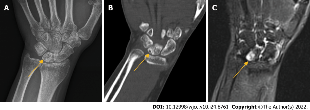
Figure 1 Preoperative imaging findings.
A-C: Preoperative radiographs (A), computed tomography (B) and magnetic resonance imaging (C), the arrow shows the necrotic lunate bone.
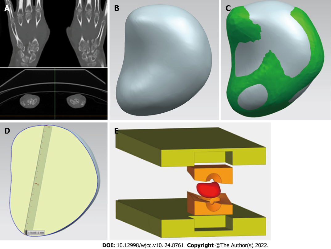
Figure 2 The process of 3D-printed injection-molded polyether ether ketone lunate prosthesis.
A: The original data of healthy lunate bone were obtained by computed tomography; B-D: 3D model of lunate prosthesis using mirroring technique; E: Prosthesis structure was prepared by injection molding.
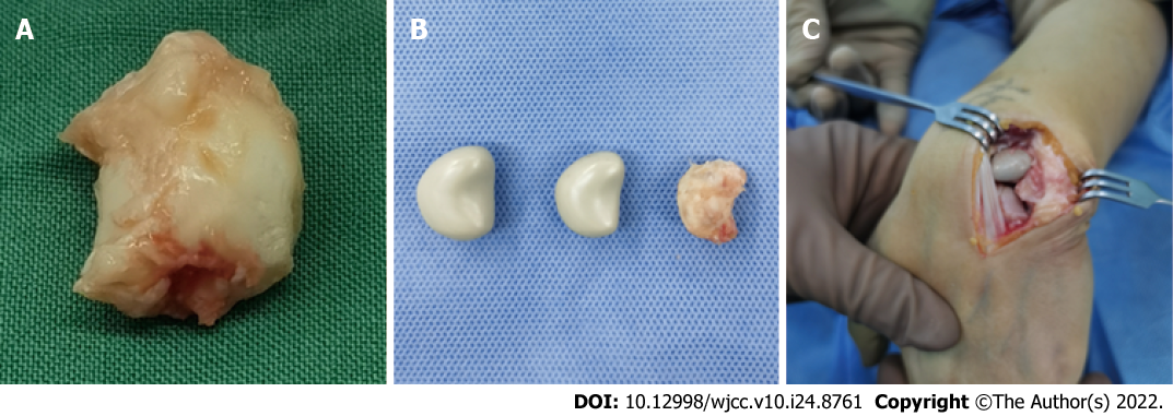
Figure 3 Intraoperative photographs.
A: Necrotic lunate bone resected; B: Necrotic lunate bone and custom-made lunate bone prosthesis of different sizes; C: Prosthesis in the original anatomic position of lunate bone.
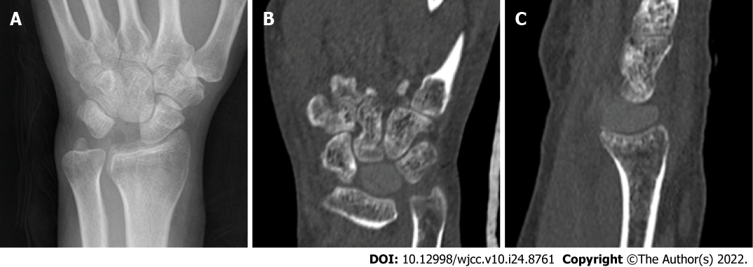
Figure 4 Postoperative imaging findings.
Postoprative radiographs (A) and computed tomography (1 yr) (B).
- Citation: Yuan CS, Tang Y, Xie HQ, Liang TT, Li HT, Tang KL. Application of 3 dimension-printed injection-molded polyether ether ketone lunate prosthesis in the treatment of stage III Kienböck’s disease: A case report. World J Clin Cases 2022; 10(24): 8761-8767
- URL: https://www.wjgnet.com/2307-8960/full/v10/i24/8761.htm
- DOI: https://dx.doi.org/10.12998/wjcc.v10.i24.8761


