Copyright
©The Author(s) 2022.
World J Clin Cases. Aug 26, 2022; 10(24): 8755-8760
Published online Aug 26, 2022. doi: 10.12998/wjcc.v10.i24.8755
Published online Aug 26, 2022. doi: 10.12998/wjcc.v10.i24.8755
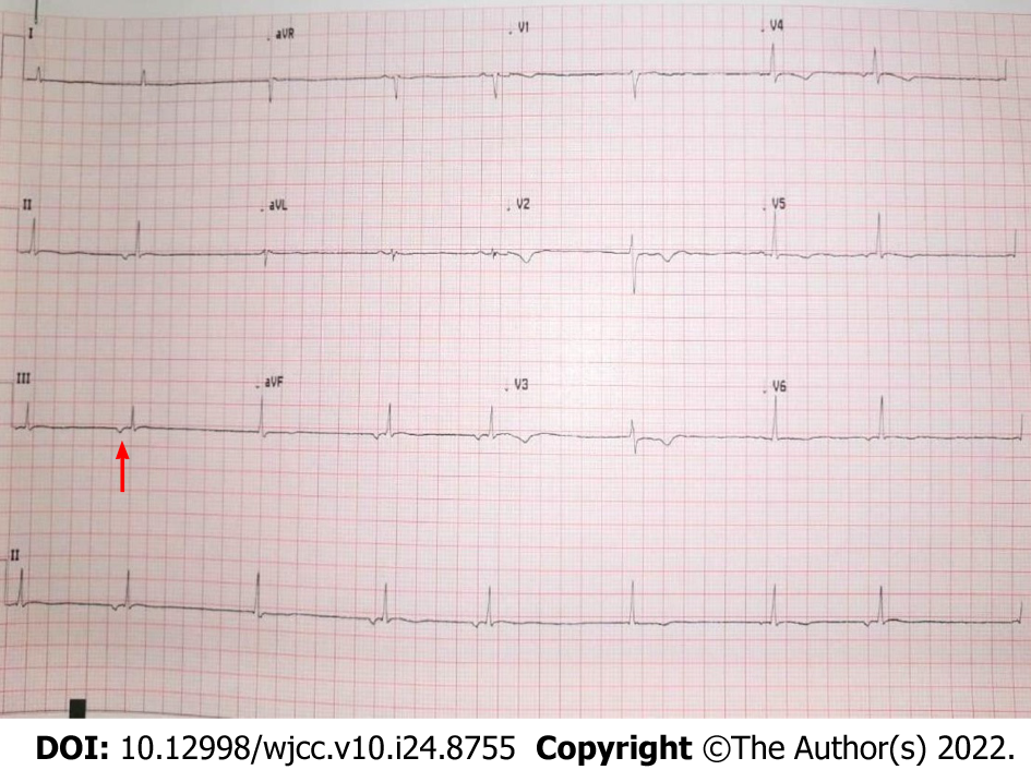
Figure 1 Patient’s electrocardiograph showing junctional bradycardia.
The inverted P wave, a key sign of junctional bradycardia, was shown by the red arrow.
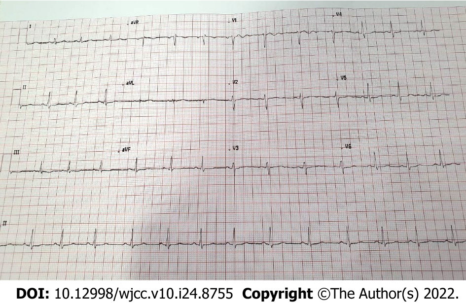
Figure 2 Patient’s electrocardiograph showing normal sinus rhythm.
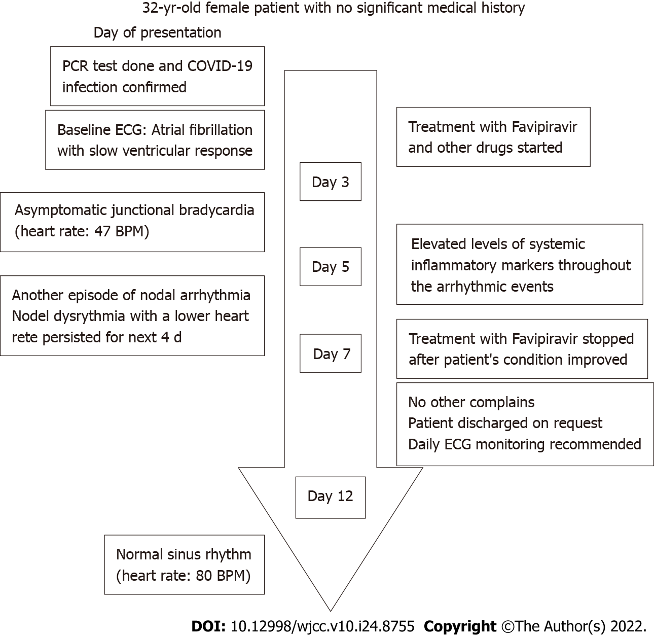
Figure 3 Patient’s flowchart.
BPM: Beats per minute; COVID-19: Coronavirus disease 2019; ECG: Electrocardiograph; PCR: Polymerase chain reaction.
- Citation: Aedh AI. Junctional bradycardia in a patient with COVID-19: A case report. World J Clin Cases 2022; 10(24): 8755-8760
- URL: https://www.wjgnet.com/2307-8960/full/v10/i24/8755.htm
- DOI: https://dx.doi.org/10.12998/wjcc.v10.i24.8755


