Copyright
©The Author(s) 2022.
World J Clin Cases. Aug 26, 2022; 10(24): 8695-8702
Published online Aug 26, 2022. doi: 10.12998/wjcc.v10.i24.8695
Published online Aug 26, 2022. doi: 10.12998/wjcc.v10.i24.8695
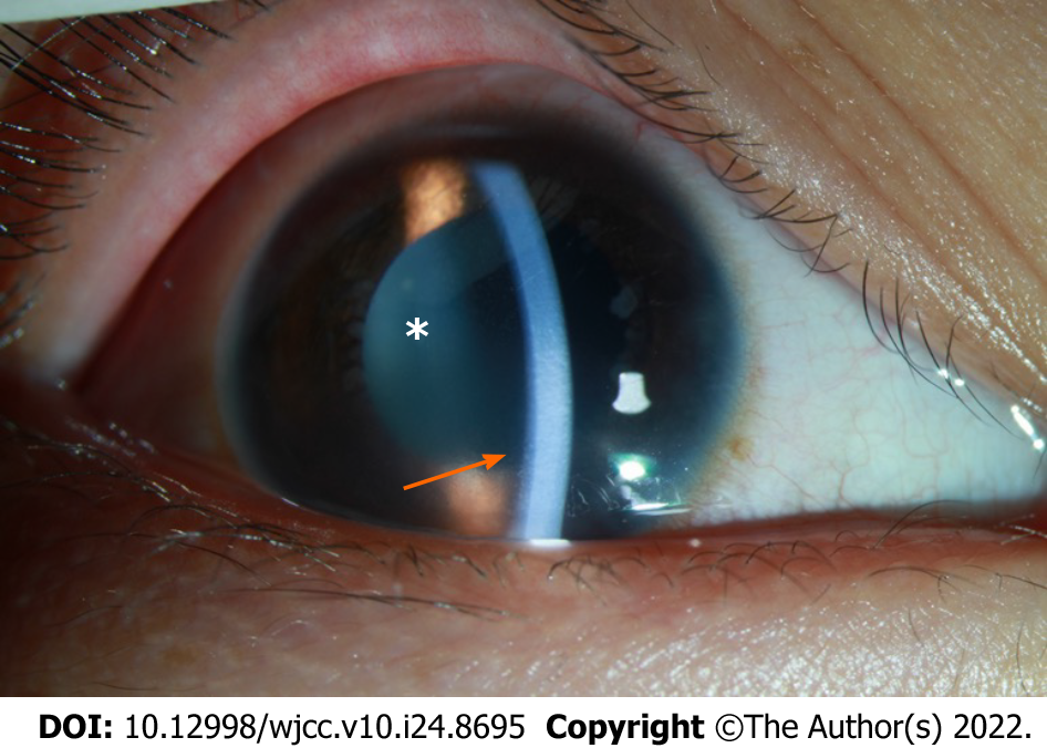
Figure 1 Image of the anterior chamber in the right eye on Day 1 after hospitalization.
The arrow shows corneal edema with haze. The depth of the anterior chamber was normal. Mutton-fat KP: ++ (arrow); anterior chamber cells: +++ (asterisk).
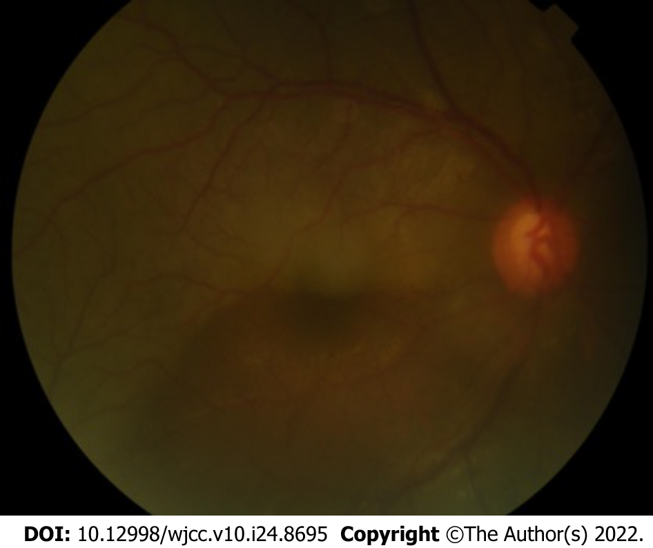
Figure 2 Color image of the fundus of the right eye.
The vitreous body was opaque, the C/D ratio of the optic disk was 0.55, retinal edema with bluish color, and the superior temporal retina was pale with occluded, line-like vessels.
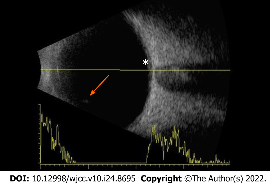
Figure 3 B-ultrasound image of the right eye.
The images show the opacity of the vitreous body (arrow) and retinal edema (asterisk).
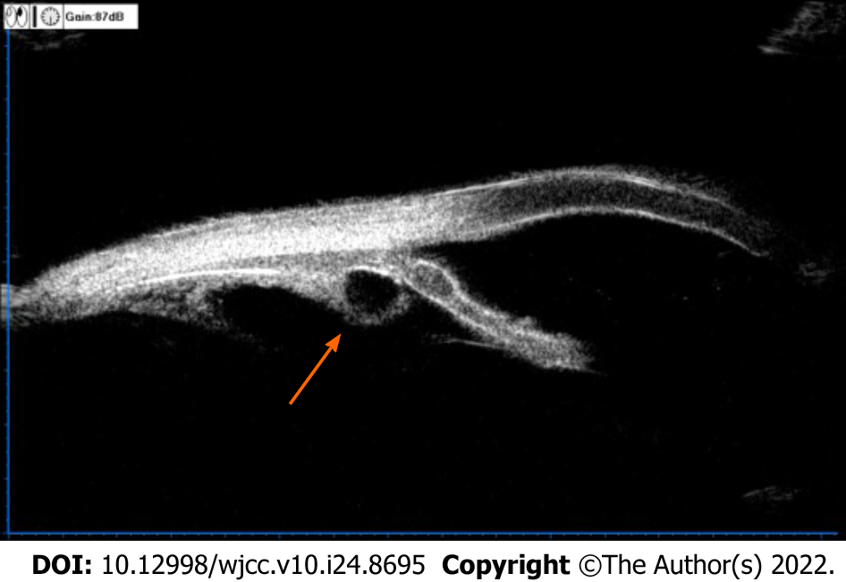
Figure 4 Image of ultrasound biomicroscopy examination of the right eye.
The result suggested multiple ciliary body cysts (arrow).
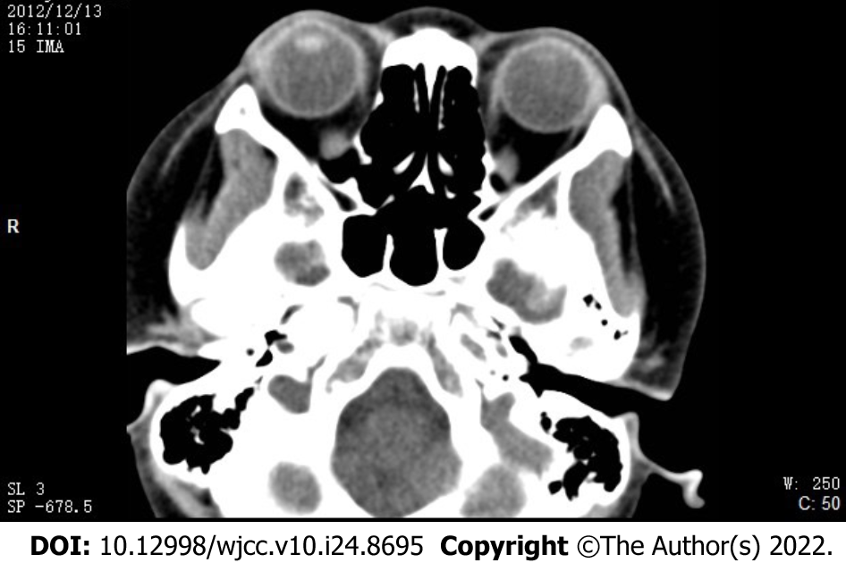
Figure 5 Computerized tomography image from axial view and coronal view.
No intraocular, space-occupying lesions were found.
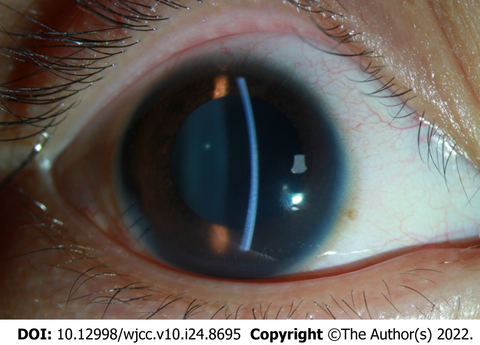
Figure 6 Image of the anterior segment of the right eye on Day 1 after the anterior chamber irrigation procedure.
The cornea was clear. The depth of the anterior chamber was normal. KP: +; AR: +.
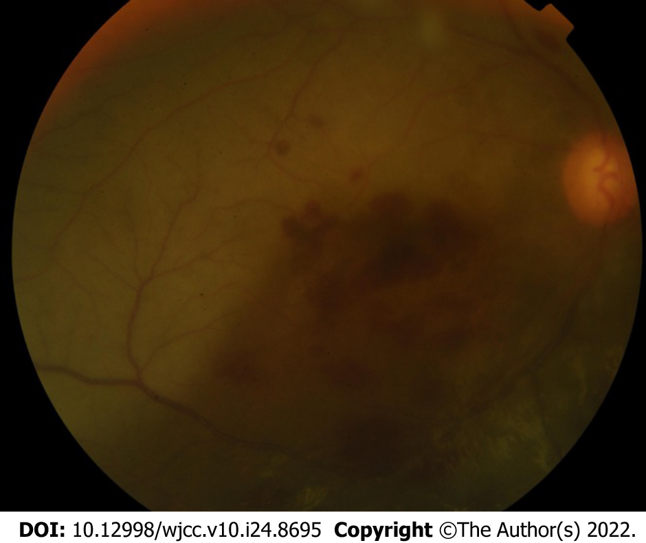
Figure 7 Color image of the fundus of the right eye after the anterior chamber irrigation procedure.
The image showed vitreous opacity, retinal edema with bluish color, and sporadic bleeding in the macular and subretinal area.
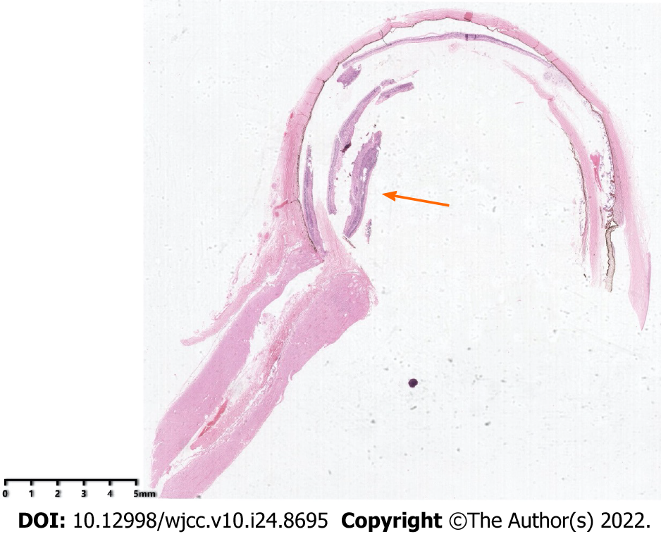
Figure 8 H&E staining of the enucleated eye.
Arrow: Tumor mass.
- Citation: Zhang Y, Tang L. Retinoblastoma in an older child with secondary glaucoma as the first clinical presenting symptom: A case report. World J Clin Cases 2022; 10(24): 8695-8702
- URL: https://www.wjgnet.com/2307-8960/full/v10/i24/8695.htm
- DOI: https://dx.doi.org/10.12998/wjcc.v10.i24.8695


