Copyright
©The Author(s) 2022.
World J Clin Cases. Aug 26, 2022; 10(24): 8686-8694
Published online Aug 26, 2022. doi: 10.12998/wjcc.v10.i24.8686
Published online Aug 26, 2022. doi: 10.12998/wjcc.v10.i24.8686
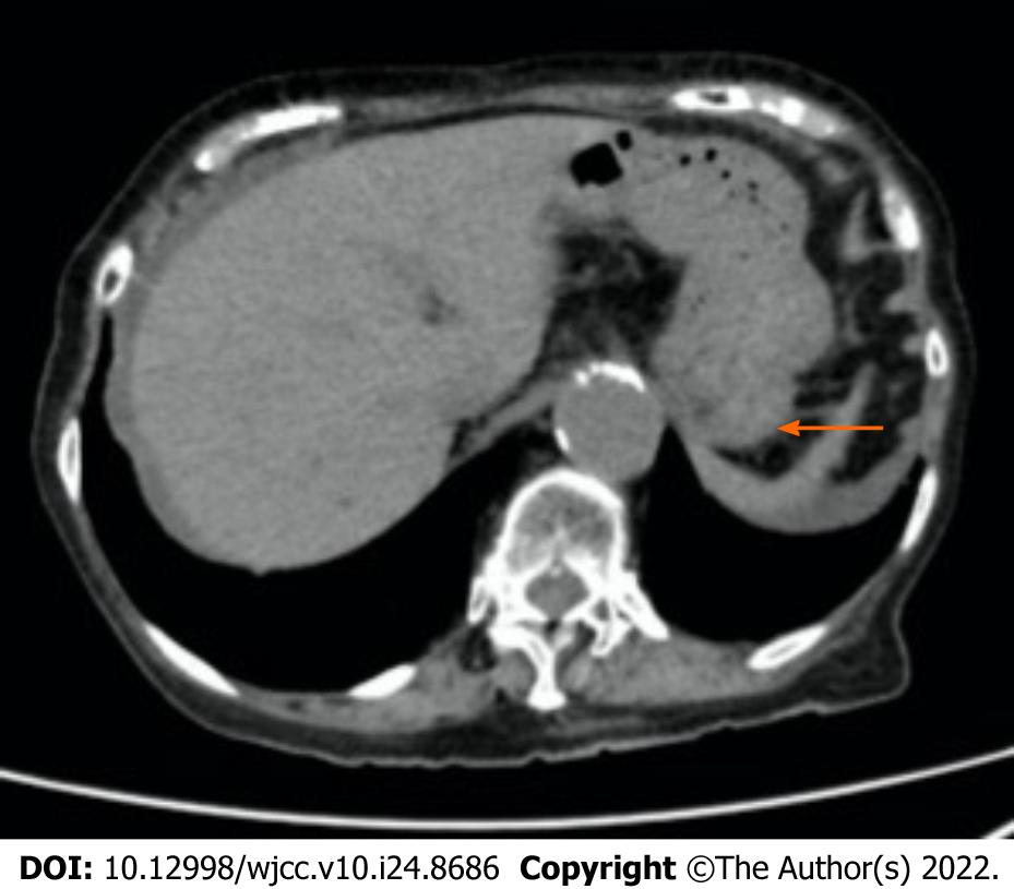
Figure 1 Abdominal plane computed tomography scans obtained 14 d before the onset of ischemic gastritis in case 1.
Computed tomography revealed wall thickening, mural emphysema, and fluid retention in the stomach. The arrow shows the wall thickening. The arrowhead indicates the mural emphysema.
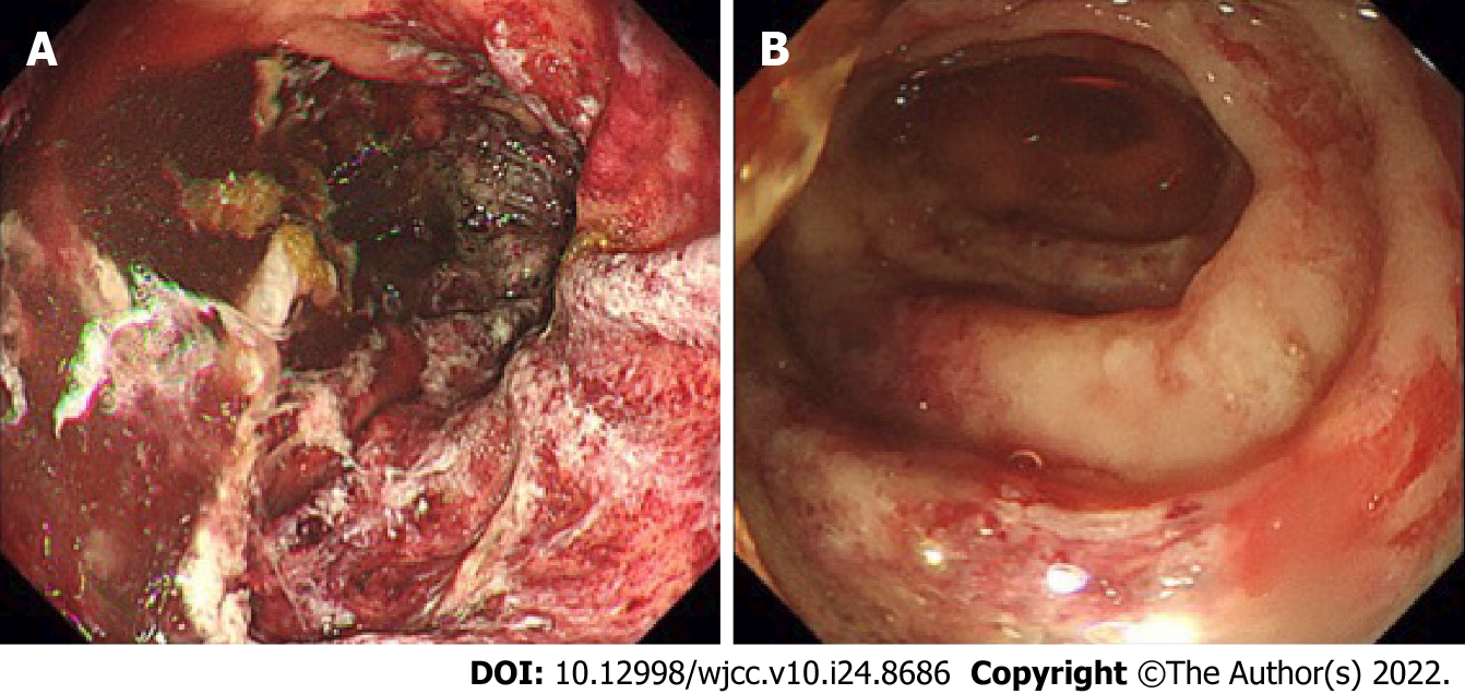
Figure 2 Upper gastrointestinal endoscopy images performed on hospital day 2 of case 1.
A: It showed longitudinal ulcers, multiple irregular ulcers, mucosal edema with redness, erosion, and hemorrhage in the stomach; B: In the duodenum, it showed longitudinal ulcers, multiple irregular ulcers, mucosal edema with redness, erosion, and hemorrhage.
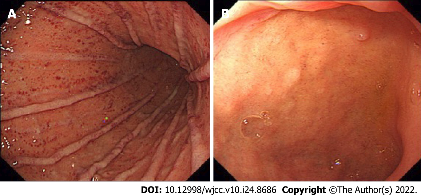
Figure 3 Upper gastrointestinal endoscopy images on hospital day 16 of case 1.
A: It revealed improved mucosal findings in the stomach; B: It revealed improved mucosal findings in the duodenum.
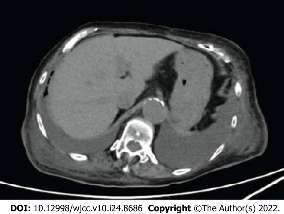
Figure 4 Abdominal plane computed tomography scans obtained after improvement of endoscopic findings.
It revealed persistent wall thickening and mural edema and significant bilateral pleural effusion.
- Citation: Shionoya K, Sasaki A, Moriya H, Kimura K, Nishino T, Kubota J, Sumida C, Tasaki J, Ichita C, Makazu M, Masuda S, Koizumi K, Kawachi J, Tsukiyama T, Kako M. Clinical features and progress of ischemic gastritis with high fatalities: Seven case reports. World J Clin Cases 2022; 10(24): 8686-8694
- URL: https://www.wjgnet.com/2307-8960/full/v10/i24/8686.htm
- DOI: https://dx.doi.org/10.12998/wjcc.v10.i24.8686


