Copyright
©The Author(s) 2022.
World J Clin Cases. Aug 26, 2022; 10(24): 8679-8685
Published online Aug 26, 2022. doi: 10.12998/wjcc.v10.i24.8679
Published online Aug 26, 2022. doi: 10.12998/wjcc.v10.i24.8679
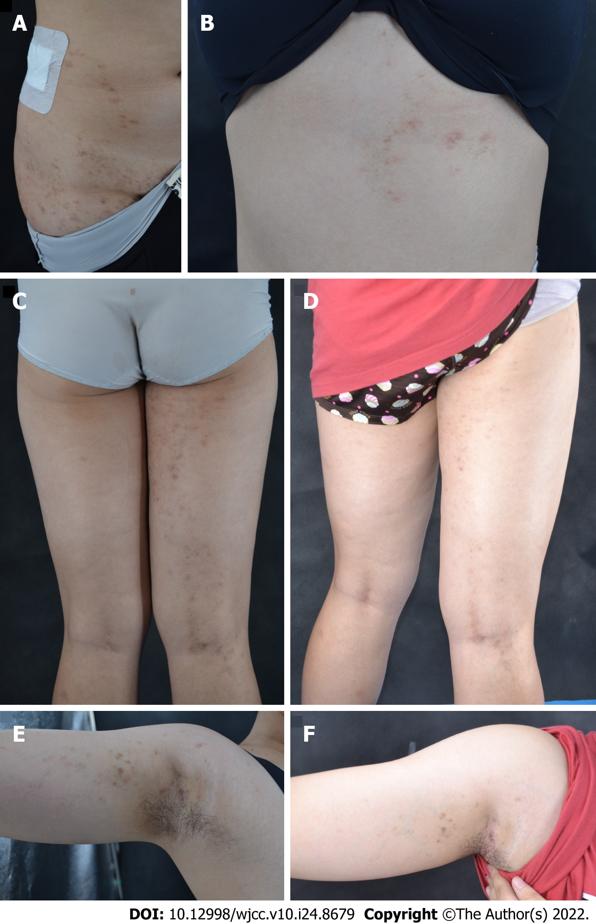
Figure 1 Clinical photographs.
A: Violaceous, brownish, polygonal papules on the right abdomen along the Blaschko’s lines; B: Violaceous, brownish papules and plaques distributed along the Blaschko’s lines on the right back and extended to the upper extremity. C: Before treatment, violaceous and red maculae with a size of 1-5 mm were seen on the right lower limb, and maculae papules were partially coalesced into patchy maculae linearly distributed; E: Before treatment, violaceous and red maculae were seen on the right upper limb; D and F: After treatment, the old lesions disappear or only pigmentation patches remain.
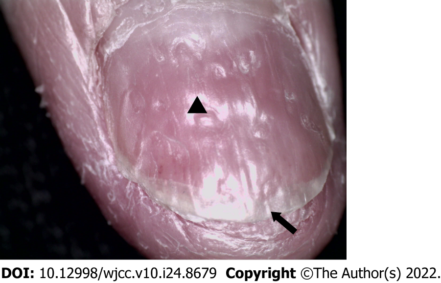
Figure 2 Nail damage.
Arrow: Distal deck splitting; Triangle: nail pits.
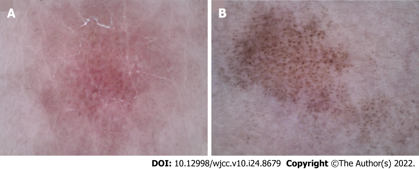
Figure 3 Dermoscopic photographs (50×).
A: Before treatment, linear and punctured vessels were seen under dermoscopy. The vascular structure was arranged radially with obvious white stripes; B: After treatment, the vascular structure disappeared, leaving blue-gray spots and faint white reticular stripes.
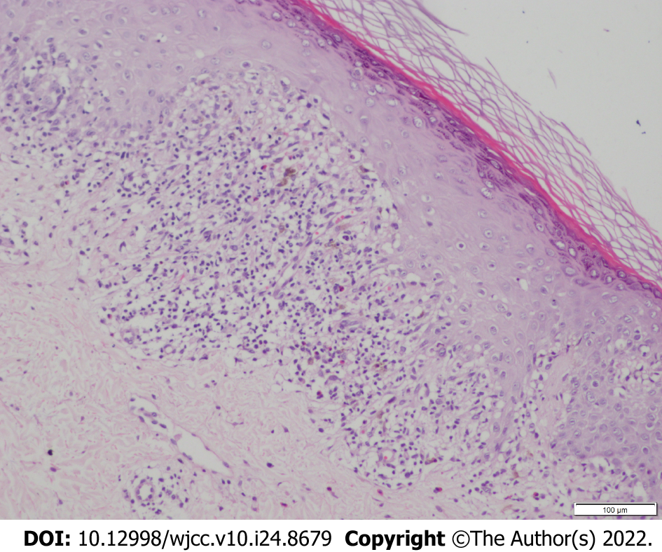
Figure 4 Skin histopathology (hematoxylin-eosin staining, 100×).
Histopathological examination showed reticular hyperkeratosis of the stratum corneum, wedge-shaped thickening of granular layer, irregular thickening of spinous layer, basal cell vacuolization and liquefaction, compact bandlike lymphocytic infiltration in superficial dermis, sporadic infiltration of chromatophilic cells, which shows typical features of lichen planus.
- Citation: Dong S, Zhu WJ, Xu M, Zhao XQ, Mou Y. Unilateral lichen planus with Blaschko line distribution: A case report. World J Clin Cases 2022; 10(24): 8679-8685
- URL: https://www.wjgnet.com/2307-8960/full/v10/i24/8679.htm
- DOI: https://dx.doi.org/10.12998/wjcc.v10.i24.8679


