Copyright
©The Author(s) 2022.
World J Clin Cases. Aug 6, 2022; 10(22): 8040-8044
Published online Aug 6, 2022. doi: 10.12998/wjcc.v10.i22.8040
Published online Aug 6, 2022. doi: 10.12998/wjcc.v10.i22.8040
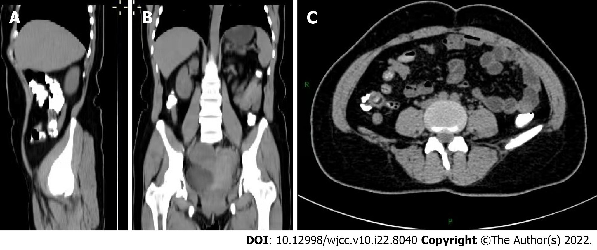
Figure 1 Abdominal computed tomography showed thickening of the appendix.
A: Median sagittal section; B: Coronal section; C: Transverse section.
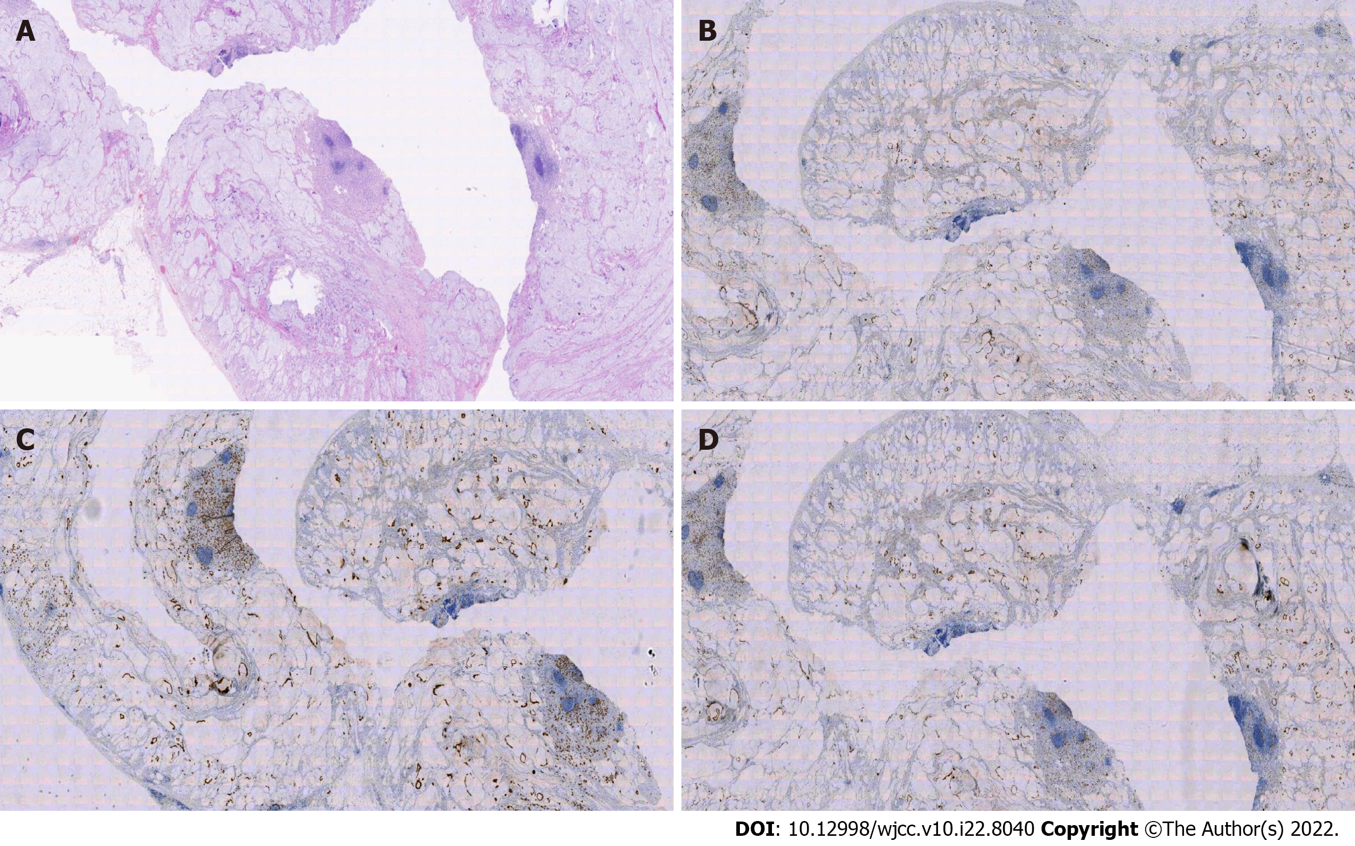
Figure 2 Histopathological examination of surgically resected specimens.
A: The cavity is filled with mucus, mucinous adenocarcinoma of the appendix, and some signet ring cell carcinoma (200×); B: Immunohistochemistry showed SATB2 (+) (200×); C: Immunohistochemistry showed CK20 (+) (200×); D: Immunohistochemistry showed CDX-2 (+) (200×).
- Citation: Wang L, Dong Y, Chen YH, Wang YN, Sun L. Accidental discovery of appendiceal carcinoma during gynecological surgery: A case report. World J Clin Cases 2022; 10(22): 8040-8044
- URL: https://www.wjgnet.com/2307-8960/full/v10/i22/8040.htm
- DOI: https://dx.doi.org/10.12998/wjcc.v10.i22.8040


