Copyright
©The Author(s) 2022.
World J Clin Cases. Aug 6, 2022; 10(22): 8034-8039
Published online Aug 6, 2022. doi: 10.12998/wjcc.v10.i22.8034
Published online Aug 6, 2022. doi: 10.12998/wjcc.v10.i22.8034
Figure 1 Initial clinical examination of the microcystic adnexal carcinoma in the local hospital.
The lesion revealed a skin-colored mass consisting of two 7 mm × 7 mm subcutaneous nodules on the glabella.
Figure 2 Three months after the surgery in our hospital.
A 30 mm × 35 mm defect was observed.
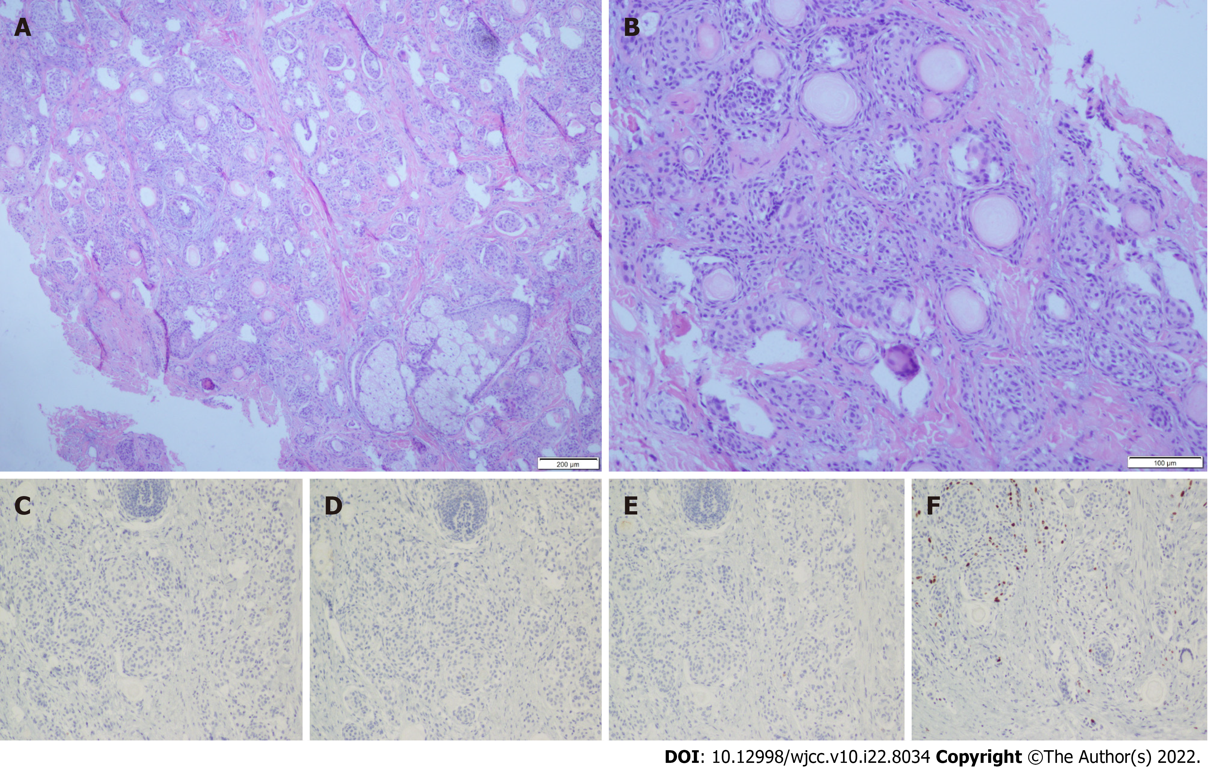
Figure 3 The histological and immunohistochemical pattern of the patient is consistent with microcystic adnexal carcinoma.
A: Original magnifications: × 40; B: Original magnifications: × 100; C: Carcinoembryonic antigen (-); D: CK7 (-); E: EMA (-); F: Ki-67(10%).
- Citation: Yang SX, Mou Y, Wang S, Hu X, Li FQ. Microcystic adnexal carcinoma misdiagnosed as a “recurrent epidermal cyst”: A case report. World J Clin Cases 2022; 10(22): 8034-8039
- URL: https://www.wjgnet.com/2307-8960/full/v10/i22/8034.htm
- DOI: https://dx.doi.org/10.12998/wjcc.v10.i22.8034


