Copyright
©The Author(s) 2022.
World J Clin Cases. Aug 6, 2022; 10(22): 7982-7988
Published online Aug 6, 2022. doi: 10.12998/wjcc.v10.i22.7982
Published online Aug 6, 2022. doi: 10.12998/wjcc.v10.i22.7982
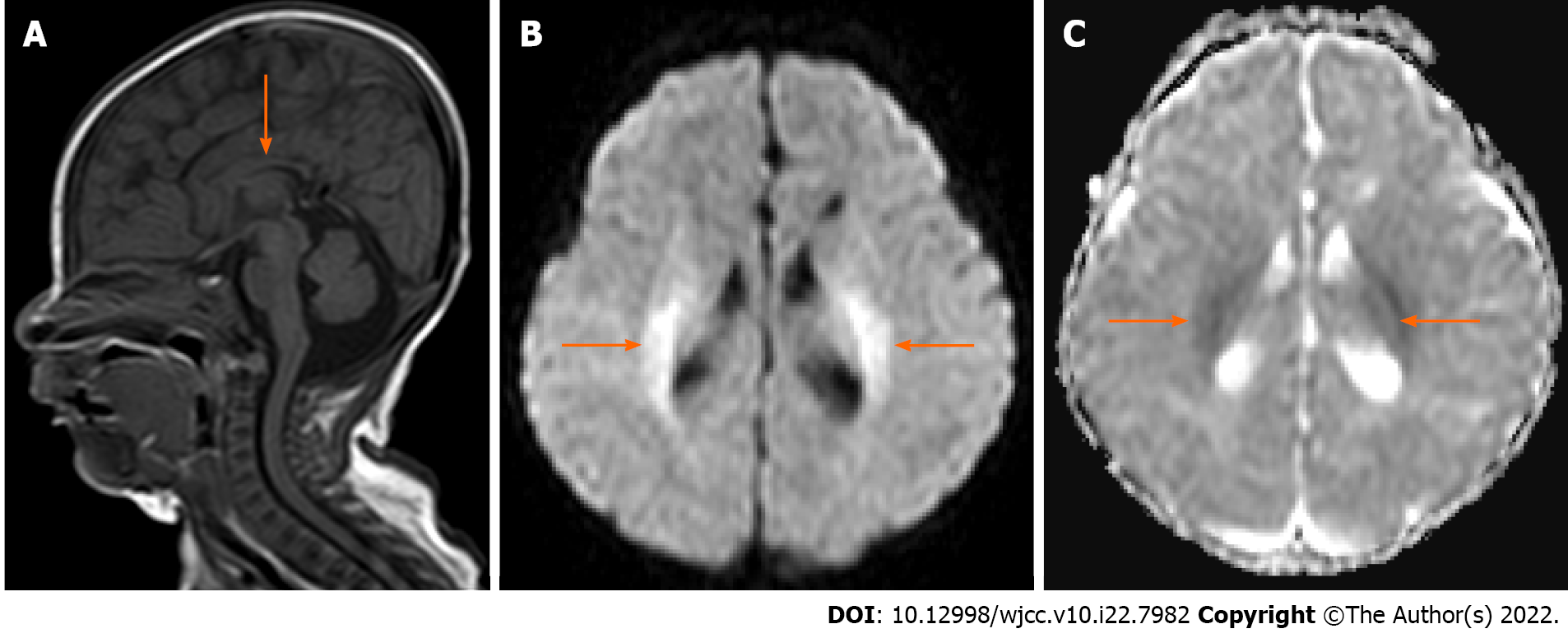
Figure 1 Head magnetic resonance imaging of the nonketotic hyperglycinemia child at the age of 2 mo.
A: The corpus callosum was small (arrow); B: On diffusion-weighted imaging, the upper corticospinal tract and bilateral paraventricular and parietal white matter showed a symmetrical high signal intensity (arrow); C: Apparent diffusion coefficient diagram showed a slightly lower signal (arrow).
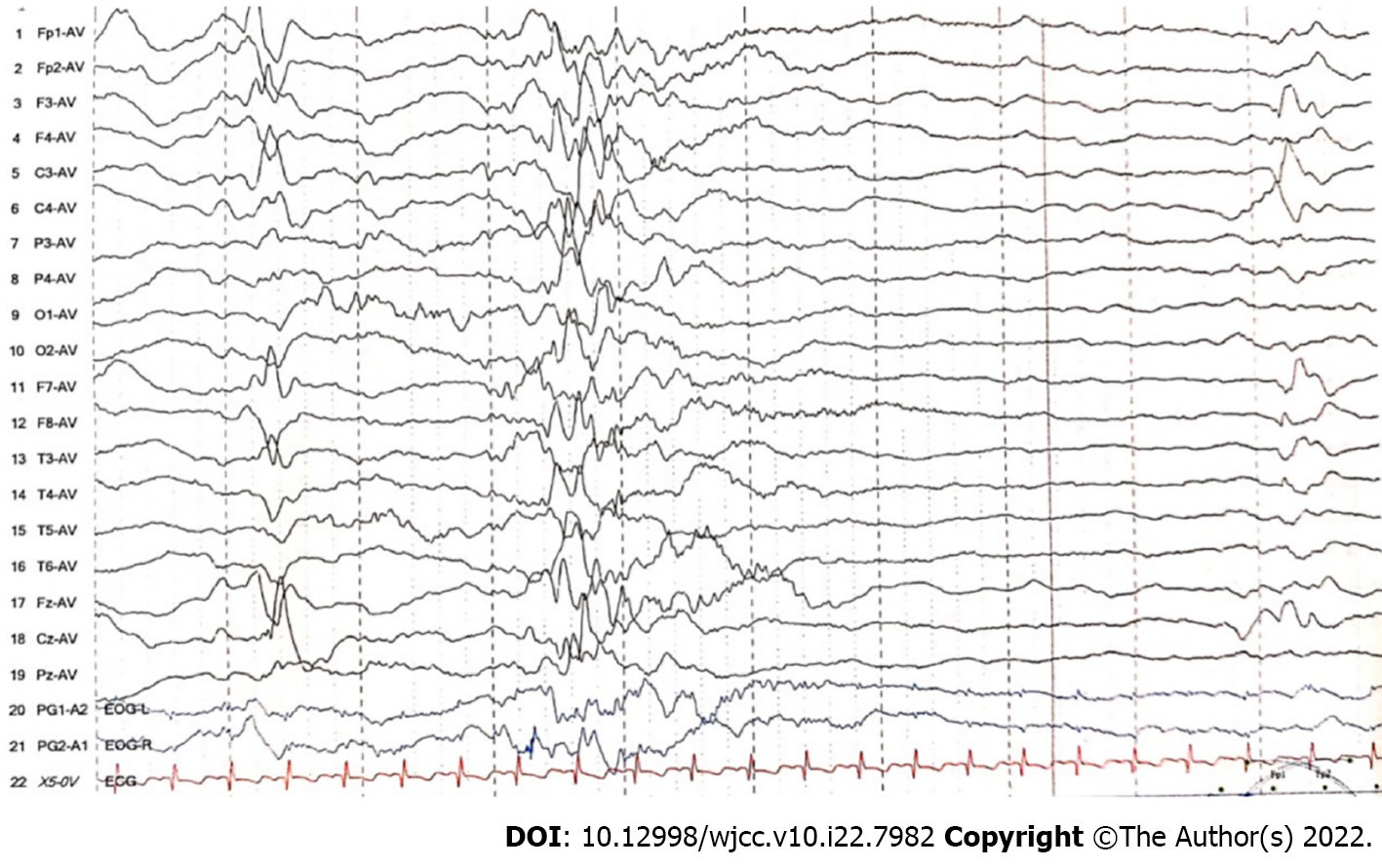
Figure 2 In the abnormal electroencephalogram, persistent multifocal or extensive irregular sharp waves or sharp slow waves could be seen during the interattack, most of which were in a burst-inhibition state, and the inhibition period lasted 2–66 s.
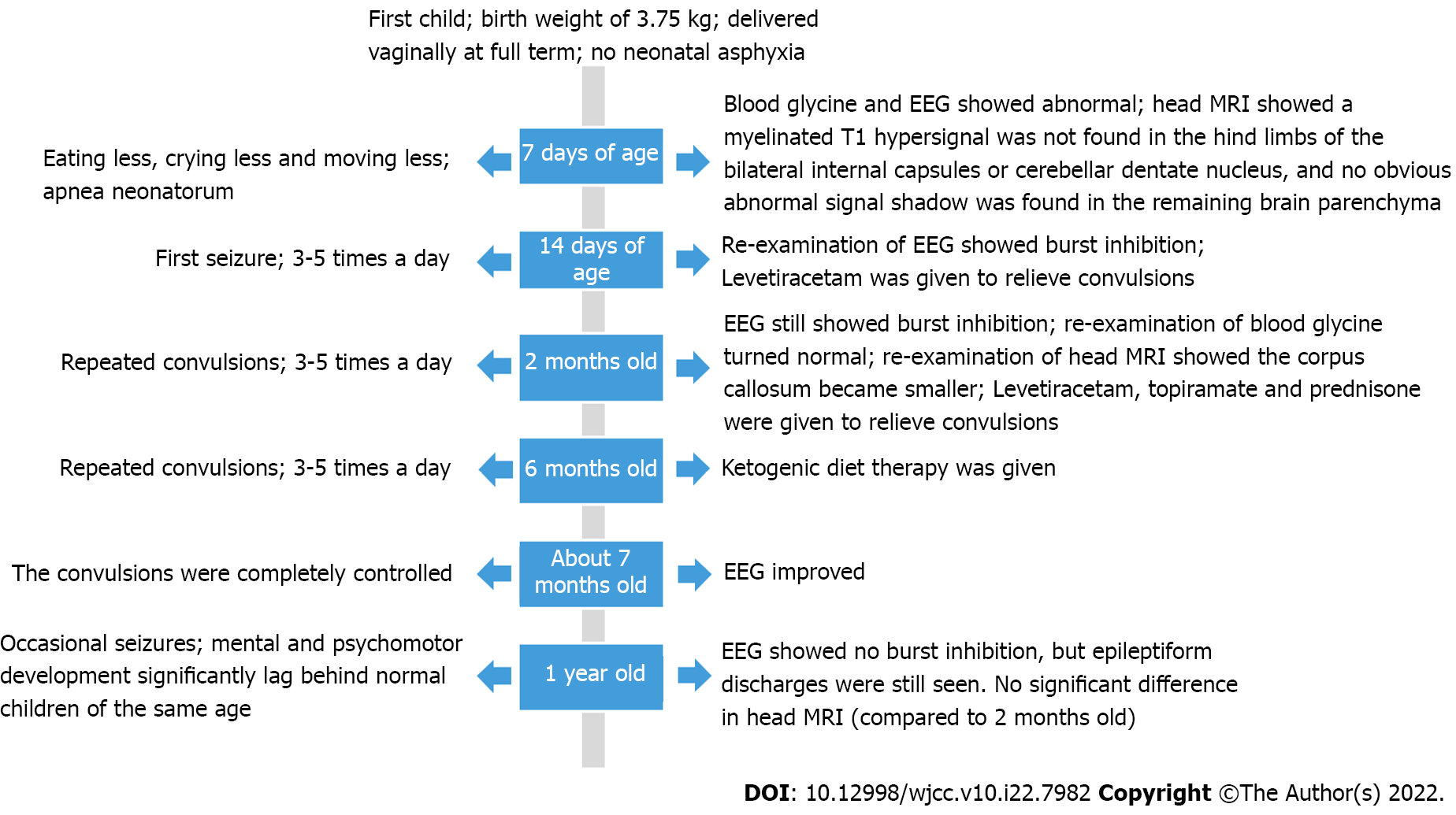
Figure 3 Time frame of main clinical information.
EEG: Electroencephalogram.
- Citation: Ning JJ, Li F, Li SQ. Clinical and genetic analysis of nonketotic hyperglycinemia: A case report. World J Clin Cases 2022; 10(22): 7982-7988
- URL: https://www.wjgnet.com/2307-8960/full/v10/i22/7982.htm
- DOI: https://dx.doi.org/10.12998/wjcc.v10.i22.7982


