Copyright
©The Author(s) 2022.
World J Clin Cases. Aug 6, 2022; 10(22): 7973-7981
Published online Aug 6, 2022. doi: 10.12998/wjcc.v10.i22.7973
Published online Aug 6, 2022. doi: 10.12998/wjcc.v10.i22.7973
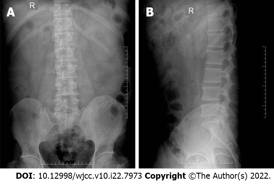
Figure 1 Pre-operative radiographs.
The intervertebral space of the 4th/5th lumbar vertebrae became smaller. A: The pre-operative anterior position radiograph; B: The pre-operative lateral position radiograph. R: Right direction.
Figure 2 Pre-operative computed tomography scans.
The position (horizontal line) of the computed tomography scan shown that there is a herniated disc of the 4th/5th lumbar vertebrae. A: The level of the 4th/5th lumbar vertebrae; B: The herniated disc of the 4th/5th lumbar vertebrae.
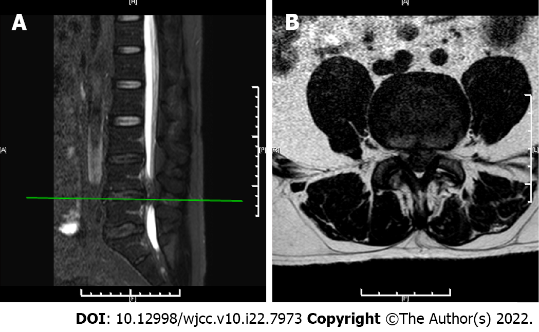
Figure 3 Pre-operative magnetic resonance imaging scans.
The position (horizontal line) of the magnetic resonance imaging scan shown that there is a herniated disc of the 4th/5th lumbar vertebrae. A: The level of the 4th/5th lumbar vertebrae; B: The herniated disc of the 4th/5th lumbar vertebrae.
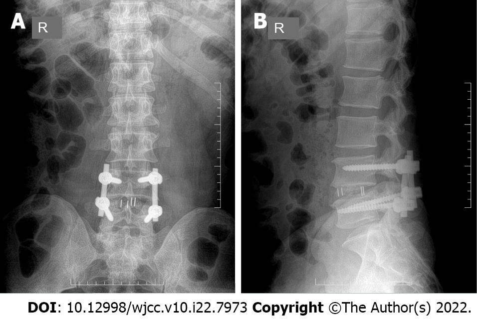
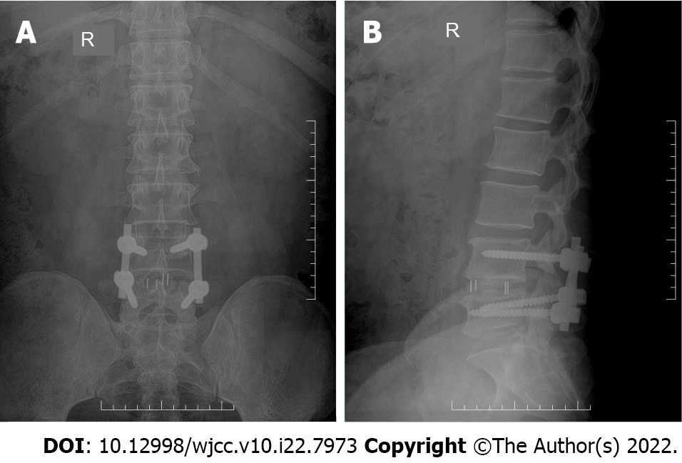
Figure 4 Post-operative radiographs.
The satisfactory positions of the lumbar spine internal fixation and fusion cage on post-operative 4-d. A: The post-operative 4-d anterior position radiograph; B: The post-operative 4-d lateral position radiograph. R: Right direction.
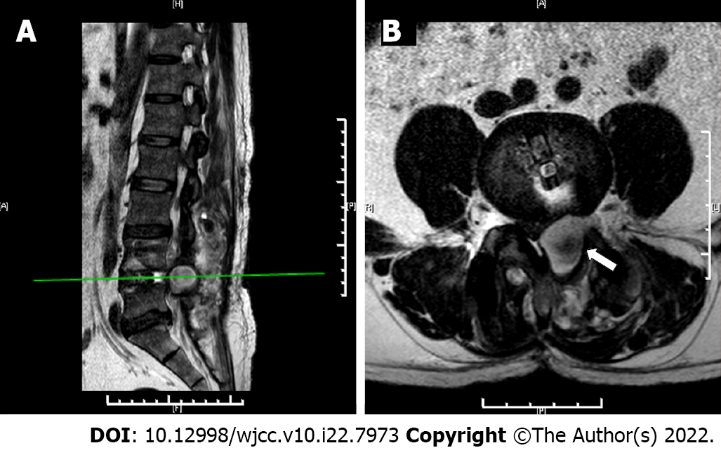
Figure 5 Post-operative magnetic resonance imaging films.
The position (horizontal line) of the magnetic resonance imaging scan shown that there is an abnormal signal in the left area of the 4th/5th lumbar vertebral body. The white arrow indicates the hematoma. A: The level of the 4th/5th lumbar vertebrae; B: The abnormal signal in the left area of the 4th/5th lumbar vertebrae.
Figure 6 The puncture treatment films.
Under C-arm X-ray machine fluoroscopy, the puncture needle in the left-side of 4th/5th lumbar vertebrae, bright red blood gushing out of the puncture needle core with continuous pulsation. A: The anterior position under C-arm X-ray machine fluoroscopy; B: The lateral position under C-arm X-ray machine fluoroscopy; C: The bright red blood gushing out of the puncture needle core with continuous pulsation.
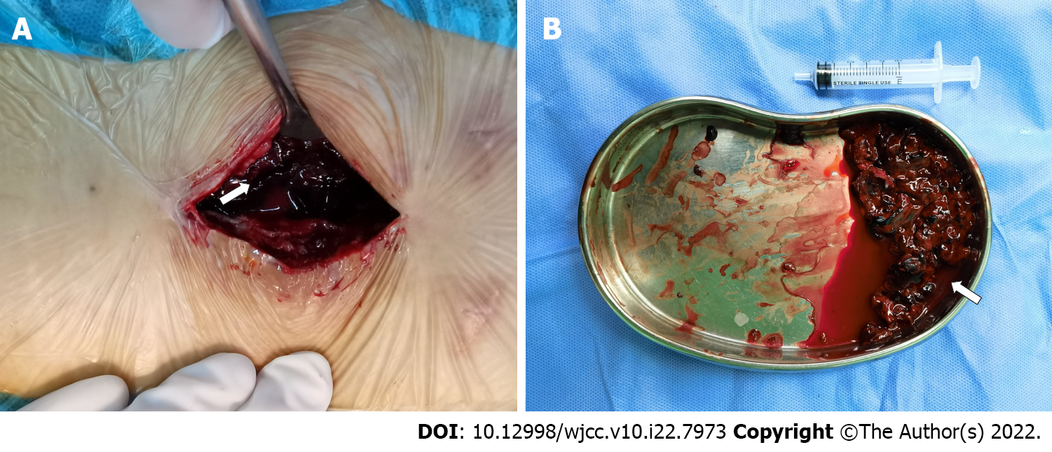
Figure 7 Surgery for hematoma removal.
The incision filled with hematoma that the removed hematoma is about 70 mL. White arrows indicate the hematoma. A: The incision filled with hematoma; B: The removed hematoma.
Figure 8 X-ray radiographs taken 9 mo post-operatively.
The satisfactory positions of the lumbar spine internal fixation and fusion cage during a 9-mo period. A: The post-operative 9-mo anterior position radiograph; B: The post-operative 9-mo lateral position radiograph. R: Right direction.
Figure 9 X-ray radiographs taken 12 mo post-operatively.
The satisfactory positions of the lumbar spine internal fixation and fusion cage during a 12-mo period. A: The post-operative 12-mo anterior position radiograph; B: The post-operative 12-mo lateral position radiograph. R: Right direction.
- Citation: Hao SS, Gao ZF, Li HK, Liu S, Dong SL, Chen HL, Zhang ZF. Delayed arterial symptomatic epidural hematoma on the 14th day after posterior lumbar interbody fusion: A case report. World J Clin Cases 2022; 10(22): 7973-7981
- URL: https://www.wjgnet.com/2307-8960/full/v10/i22/7973.htm
- DOI: https://dx.doi.org/10.12998/wjcc.v10.i22.7973


