Copyright
©The Author(s) 2022.
World J Clin Cases. Aug 6, 2022; 10(22): 7936-7943
Published online Aug 6, 2022. doi: 10.12998/wjcc.v10.i22.7936
Published online Aug 6, 2022. doi: 10.12998/wjcc.v10.i22.7936
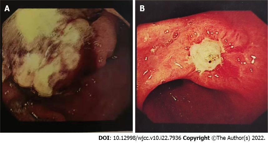
Figure 1 Upper gastrointestinal endoscopy.
A: There was a large mass in the greater curvature of the stomach with unclear borders, accompanied by ulcers (case 1); B: There was a pitted ulcer in the corner of the stomach, and the surrounding gastric mucosa was markedly congested and edematous (case 2).
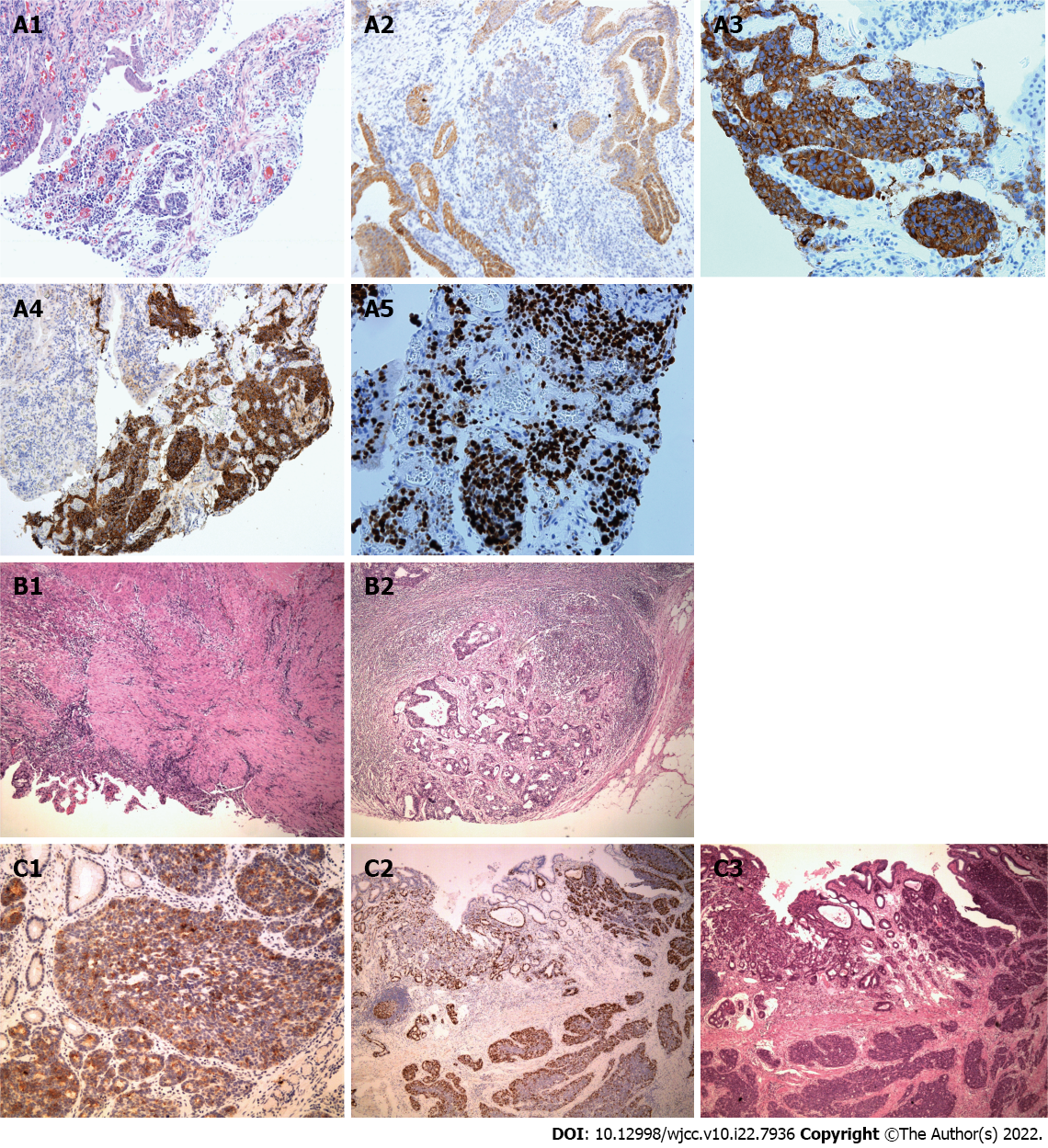
Figure 2 Immunoprofile and hematoxylin-eosin staining.
A: Immunoprofile of endoscopy specimen in case 1. A1: Hematoxylin-eosin (H&E) staining; A2-4: CK18 (A2), Syn (A3), and CgA (A4) were positive in the solid component; A5: A high Ki67 index (about 80%) can be seen; B: H&E staining of the surgery specimen in case 1. B1: A small amount of atypical glands in the muscle layer at the bottom of the ulcer area, and the surrounding fibrous tissue hyperplasia accompanied by infiltration of interstitial inflammatory cells; B2: Adenocarcinoma metastases can be seen in surrounding lymph nodes; C: Immunoprofile of surgical specimen in case 2. C1: The neuroendocrine carcinoma component was positive for CgA; C2: A high Ki67 proliferation index (about 60%) can be seen; C3: Neuroendocrine carcinoma component was dominant (about 80%).
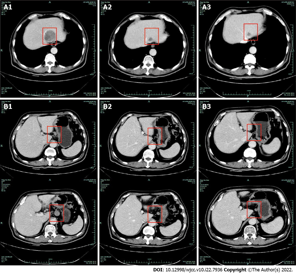
Figure 3 Computed tomography images of case 1.
A: Metastasis of the left liver. A1: At the baseline; A2: After the 2nd cycle of chemotherapy; A3: After the 3rd cycle of chemotherapy; B: Surrounding lymph nodes. B1: At the baseline, B2: After the 2nd cycle of chemotherapy; B3: After the 3rd cycle of chemotherapy.
- Citation: Woo LT, Ding YF, Mao CY, Qian J, Zhang XM, Xu N. Long-term survival of gastric mixed neuroendocrine-non-neuroendocrine neoplasm: Two case reports. World J Clin Cases 2022; 10(22): 7936-7943
- URL: https://www.wjgnet.com/2307-8960/full/v10/i22/7936.htm
- DOI: https://dx.doi.org/10.12998/wjcc.v10.i22.7936


