Copyright
©The Author(s) 2022.
World J Clin Cases. Aug 6, 2022; 10(22): 7913-7923
Published online Aug 6, 2022. doi: 10.12998/wjcc.v10.i22.7913
Published online Aug 6, 2022. doi: 10.12998/wjcc.v10.i22.7913
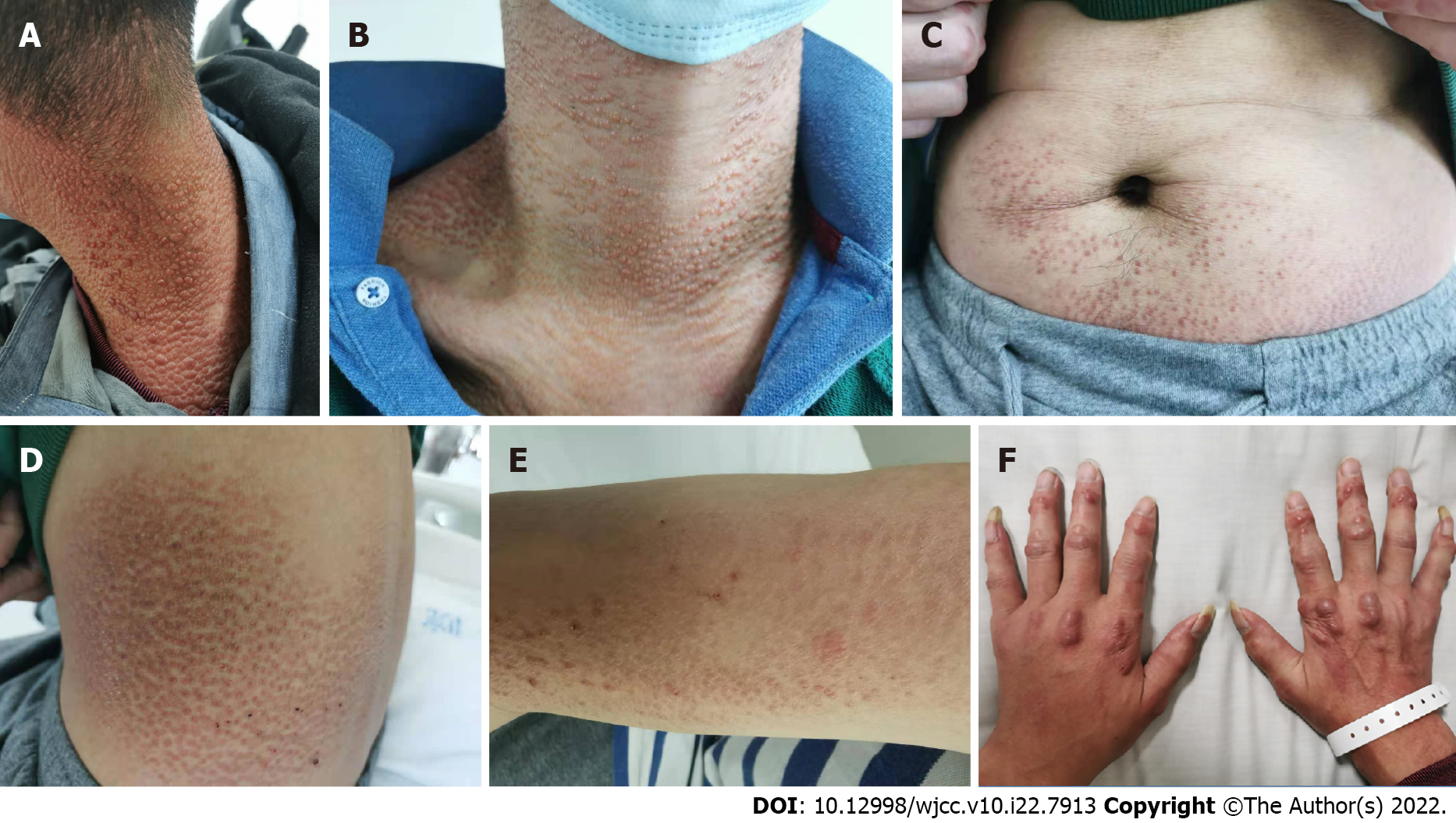
Figure 1 Skin lesions of the patient.
A-E: Skin-colored, brownish-red, millet to mung bean-sized maculopapules were observed on the patient’s neck, chest, waist, abdomen, and arms; F: Multiple round or oval nodules, 2-8 mm in size, reddish-brown, hard, and unbroken were observed on both hands.
Figure 2 Imaging examinations of the patient.
A: Radiographs of both hands. Narrowing of the interphalangeal and intercarpal joint spaces in both hands, reduced bone density of the joint components, visible cystic translucent areas, and narrowing of the radial carpal joint on the left side were observed; B: Knee MRI. Osteochondral damage of the patellofemoral articular surface and fluid accumulation in the left knee joint cavity and suprapatellar capsule were observed; C: HRCT of the chest. There was interstitial inflammation present in both lungs; bilateral interlobular fissure nodules; focal fibrosis in both lower lobes of the lungs; and cystic foci in the right subscapularis muscle.
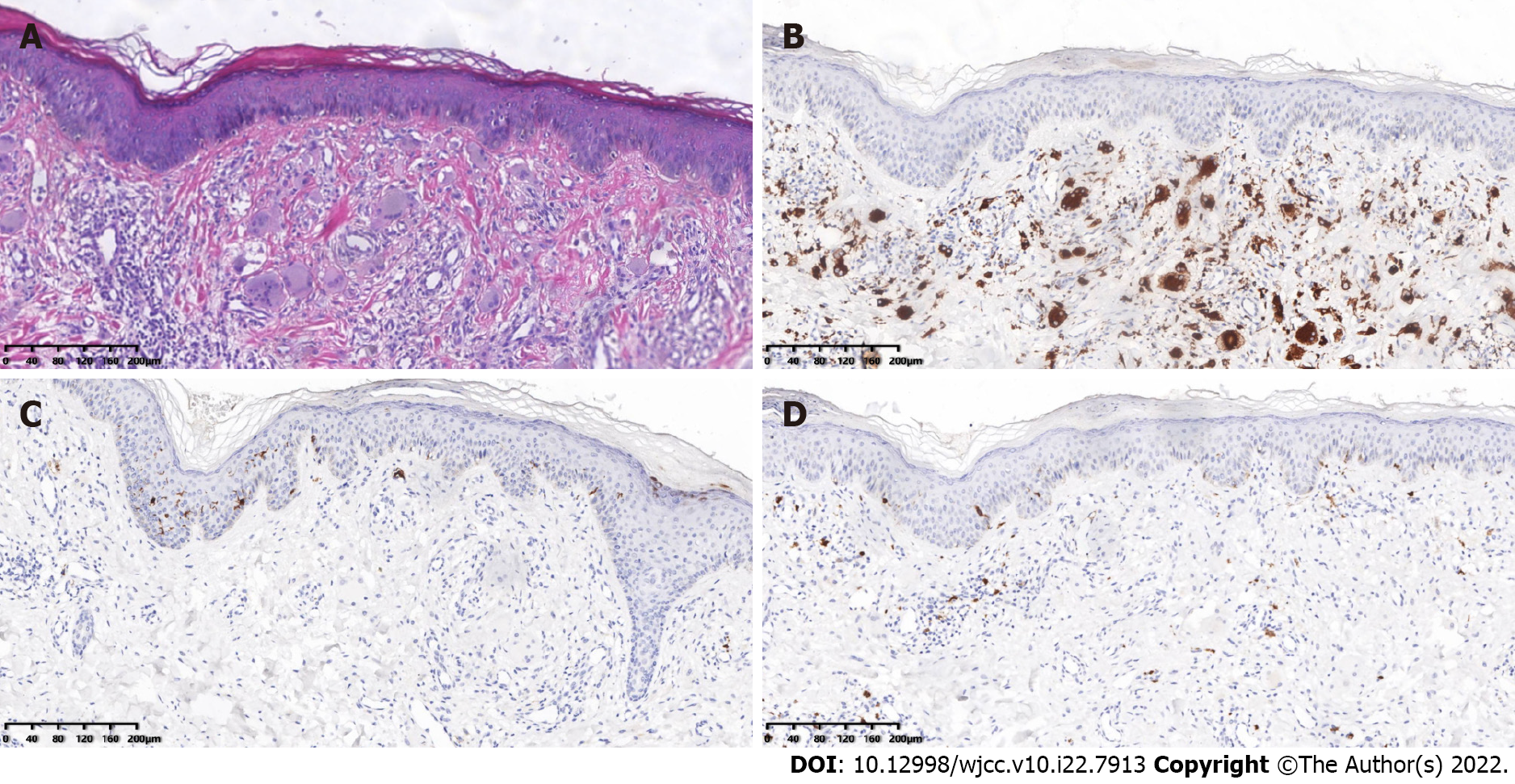
Figure 3 Pathological examination of the patient’s skin.
A: Histopathology of the skin from the left upper back. The epidermis was generally normal, and the dermal papillae showed an increased number of histiocytes and multinucleated giant cells with abundant cell cytoplasm and hairy glass-like changes, with a little surrounding lymphatic and eosinophilic infiltration (HE 40×). Immunohistochemical staining: B: CD68-positive; C: CD1a negative; and D: S-100 negative (SP×100).
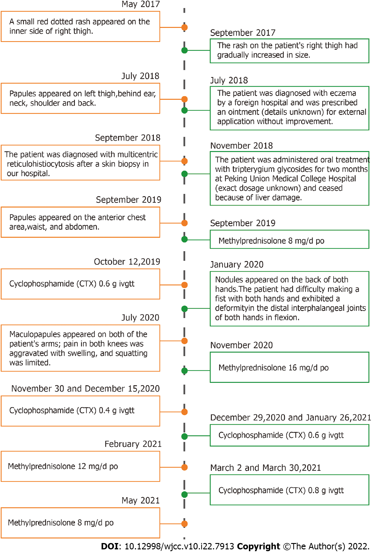
Figure 4 Timeline summarizing the patient’s disease process.
- Citation: Xu XL, Liang XH, Liu J, Deng X, Zhang L, Wang ZG. Multicentric reticulohistiocytosis with prominent skin lesions and arthritis: A case report. World J Clin Cases 2022; 10(22): 7913-7923
- URL: https://www.wjgnet.com/2307-8960/full/v10/i22/7913.htm
- DOI: https://dx.doi.org/10.12998/wjcc.v10.i22.7913


