Copyright
©The Author(s) 2022.
World J Clin Cases. Jul 26, 2022; 10(21): 7565-7570
Published online Jul 26, 2022. doi: 10.12998/wjcc.v10.i21.7565
Published online Jul 26, 2022. doi: 10.12998/wjcc.v10.i21.7565
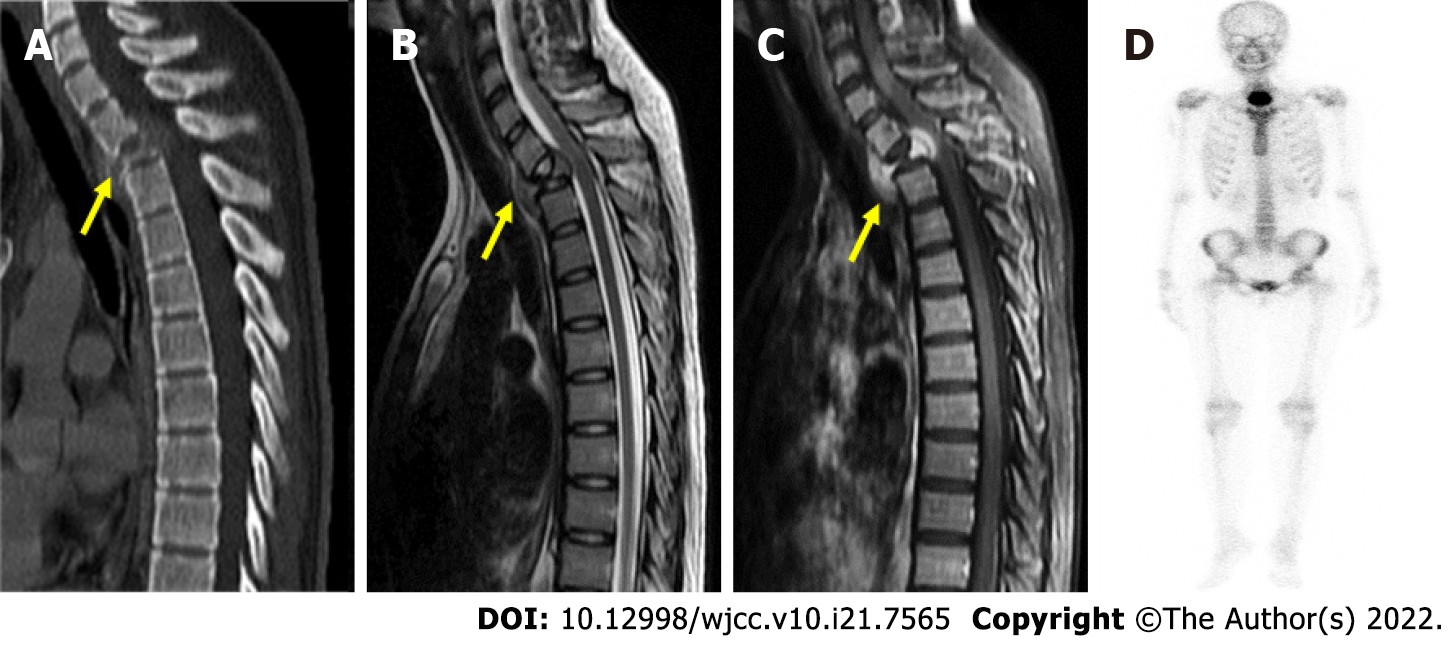
Figure 1 Preoperative radiographic evaluation.
A: Preoperative computed tomography demonstrating collapsed T2 vertebra (yellow arrow); B, C: Preoperative magnetic resonance imaging revealed vertebral body mass (yellow arrow) with paraspinal and epidural paraspinal extension and gadolinium enhancement; D: Bone scan showing an active bone lesion at the upper thoracic spine.
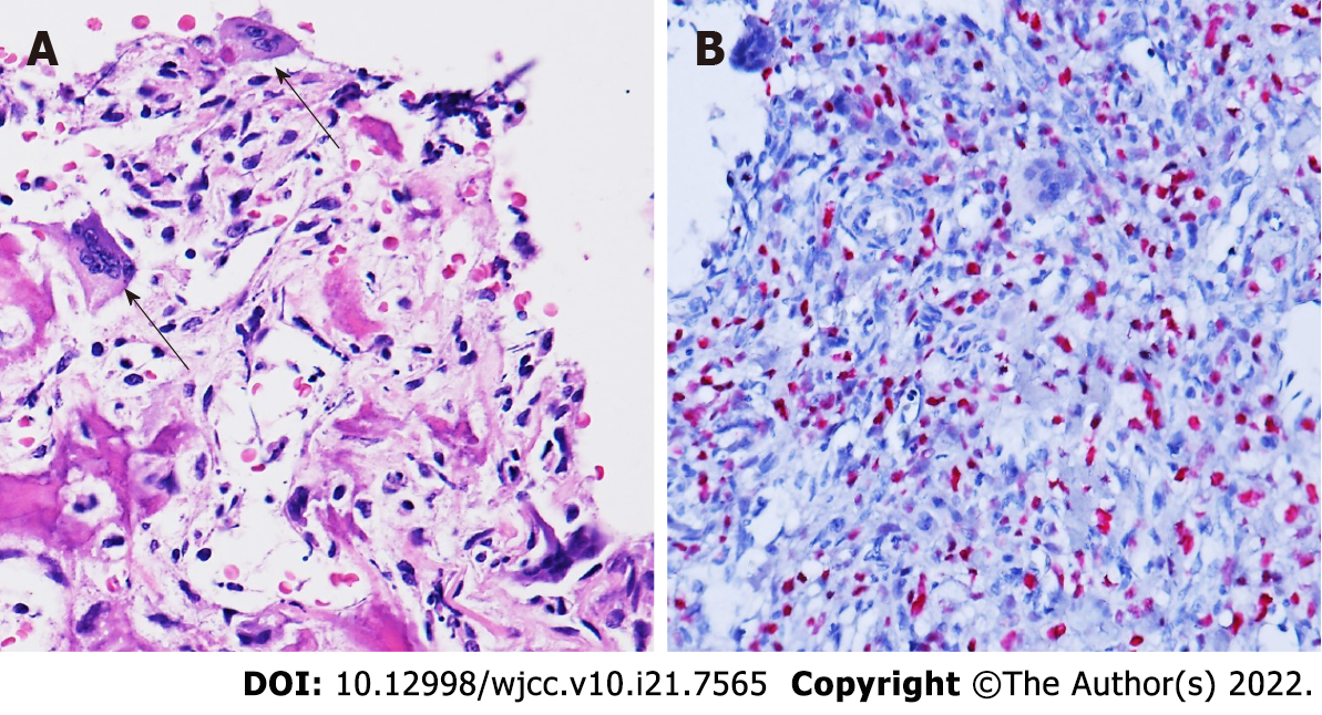
Figure 2 Histopathology of the T2 vertebral tumor.
A: Hematoxylin and eosin staining revealing atypical mononuclear oval to plump spindle cells with osteoid formation infiltrating osteoclast-like multinucleated giant cells (arrow); B: Immunohistochemistry demonstrating diffuse H3F3A G34W mutation. Magnification: 400 x.
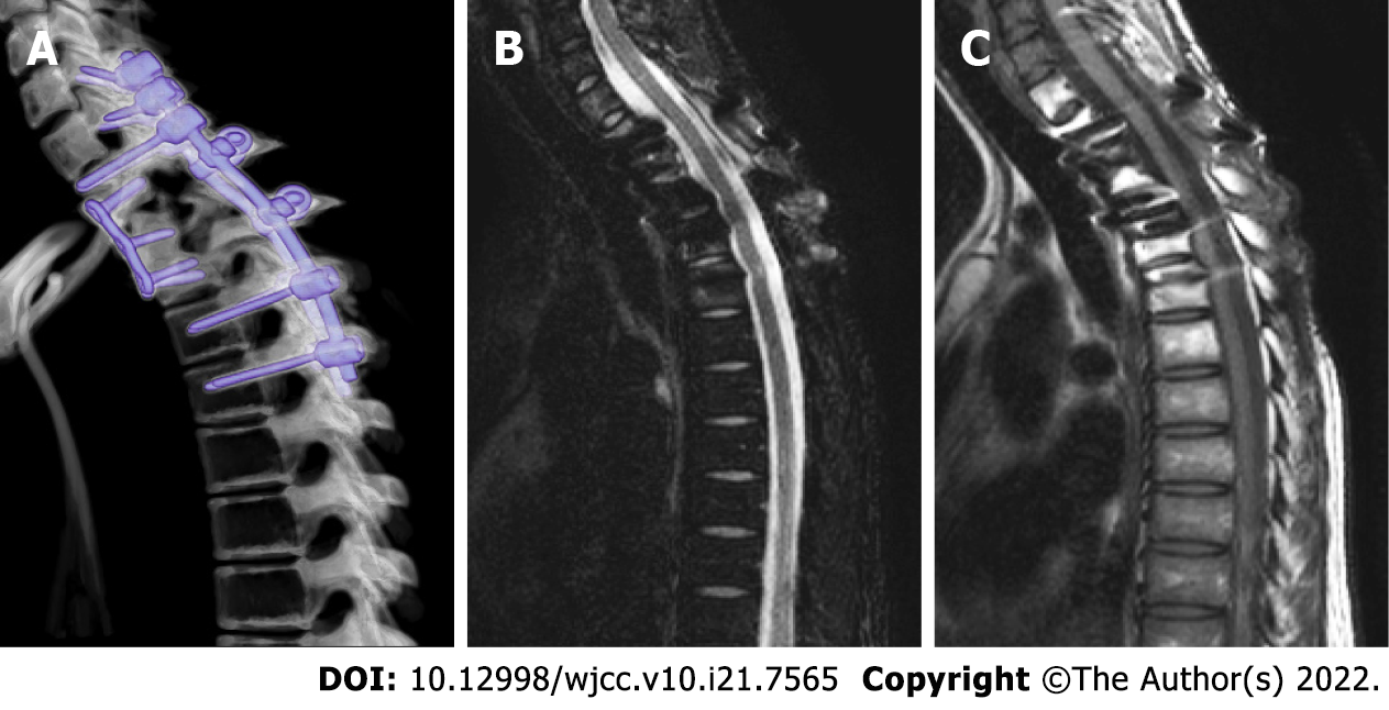
Figure 3 Postoperative radiographic evaluation.
A-C: No tumor recurrence was revealed in the A: 3-mo postoperative computed tomography; B: 4-yr postoperative T2; or C: T1 gadolinium-enhanced magnetic resonance imaging.
- Citation: Tseng CS, Wong CE, Huang CC, Hsu HH, Lee JS, Lee PH. Spinal giant cell-rich osteosarcoma-diagnostic dilemma and treatment strategy: A case report. World J Clin Cases 2022; 10(21): 7565-7570
- URL: https://www.wjgnet.com/2307-8960/full/v10/i21/7565.htm
- DOI: https://dx.doi.org/10.12998/wjcc.v10.i21.7565


