Copyright
©The Author(s) 2022.
World J Clin Cases. Jul 26, 2022; 10(21): 7553-7564
Published online Jul 26, 2022. doi: 10.12998/wjcc.v10.i21.7553
Published online Jul 26, 2022. doi: 10.12998/wjcc.v10.i21.7553
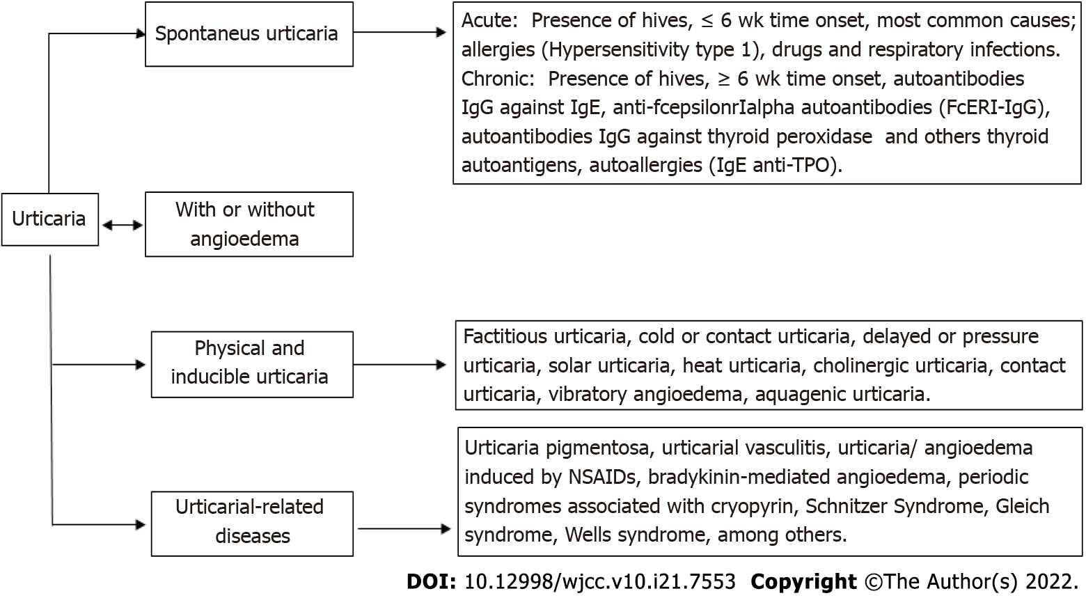
Figure 1 Classification of urticaria according to the World Allergy Organization, the European Academy of Allergy and Clinical Immunology, the Global Allergy and Asthma European Network and the European Dermatology Forum.
Figure 2 Visual representation of the different lesions found in the patient.
A: In the oropharynx, nodular lesions of approximately 0.5 cm in diameter are evidenced; B: Erythematous, wheal-like lesions with raised and serpiginous edges are observed, highly pruritic, with intralesional confluence on the anterior trunk and upper extremities; C: Lesions with similar characteristics on the posterior trunk. Images were taken under patient´s consent.
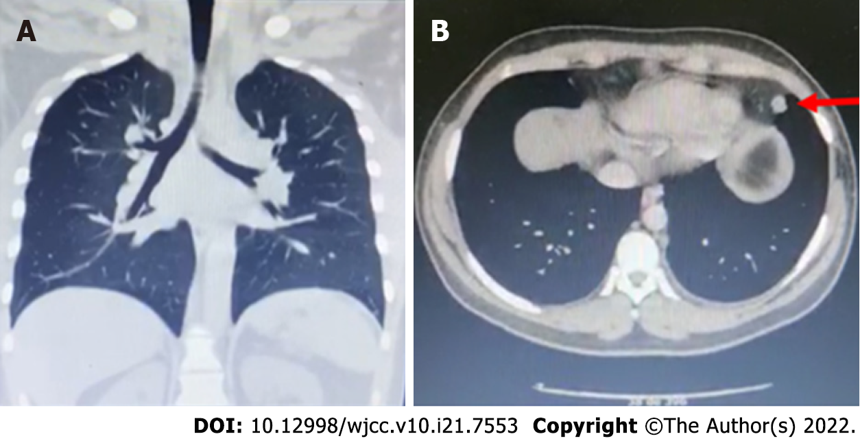
Figure 3 Chest computed tomography.
A: Chest computed tomography coronal section showing the presence of paratracheal nodules; B: Larger nodule at the level of the lingula in axial section.
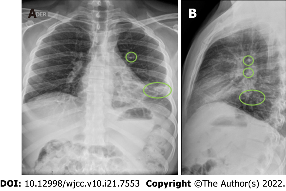
Figure 4 Chest X-ray findings.
A, B: Anteroposterior and lateral chest X-ray. Multiple nodules are observed in both pulmonary fields, predominantly perihilar.
- Citation: Jiménez LF, Castellón EA, Marenco JD, Mejía JM, Rojas CA, Jiménez FT, Coronell L, Osorio-Llanes E, Mendoza-Torres E. Chronic urticaria associated with lung adenocarcinoma — a paraneoplastic manifestation: A case report and literature review . World J Clin Cases 2022; 10(21): 7553-7564
- URL: https://www.wjgnet.com/2307-8960/full/v10/i21/7553.htm
- DOI: https://dx.doi.org/10.12998/wjcc.v10.i21.7553


