Copyright
©The Author(s) 2022.
World J Clin Cases. Jul 26, 2022; 10(21): 7429-7437
Published online Jul 26, 2022. doi: 10.12998/wjcc.v10.i21.7429
Published online Jul 26, 2022. doi: 10.12998/wjcc.v10.i21.7429
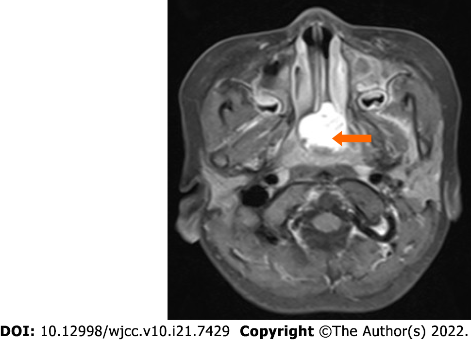
Figure 1 Enhanced magnetic resonance imaging showing an abnormal signal in the nasopharynx with obvious enhancement.
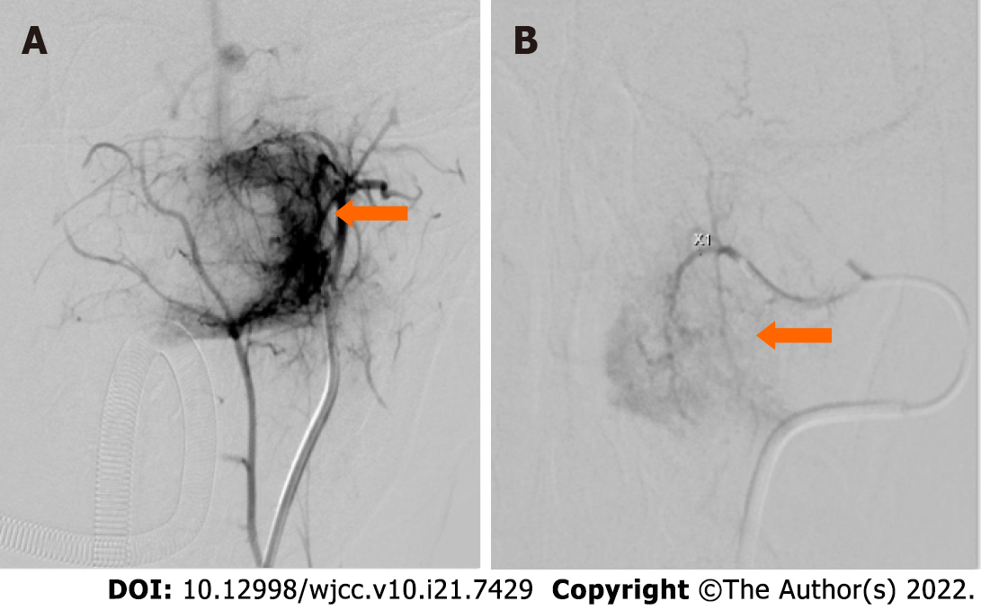
Figure 2 Before and after embolization.
A: Pre-embolization angiography reveals that the tumor (arrow) is supplied by the left internal maxillary artery; B: After the second embolization, the arteriovenous fistula in the tumor in the nasopharynx has almost disappeared.
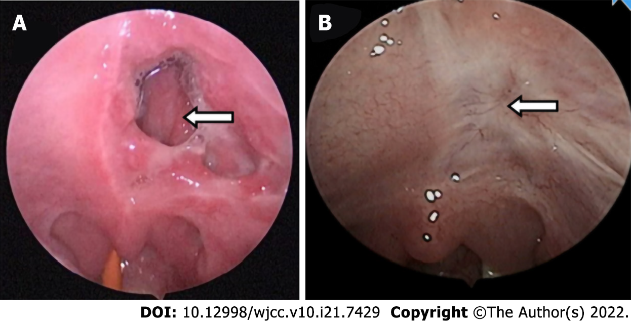
Figure 3 Transformation of soft palate perforation.
A: After the first embolization, an irregular perforation is seen in the left soft palate; B: At 3 mo after the endoscopic excision, the necrotic area in the left soft palate has completely healed.
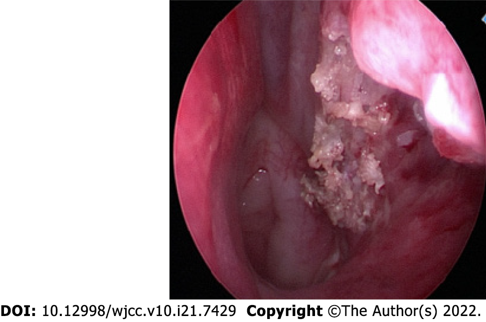
Figure 4 The posterior end of the inferior turbinate and lateral wall of the nasopharynx after resection of the tumor mass.
- Citation: Yan YY, Lai C, Wu L, Fu Y. Extranasopharyngeal angiofibroma in children: A case report. World J Clin Cases 2022; 10(21): 7429-7437
- URL: https://www.wjgnet.com/2307-8960/full/v10/i21/7429.htm
- DOI: https://dx.doi.org/10.12998/wjcc.v10.i21.7429


