Copyright
©The Author(s) 2022.
World J Clin Cases. Jul 26, 2022; 10(21): 7356-7364
Published online Jul 26, 2022. doi: 10.12998/wjcc.v10.i21.7356
Published online Jul 26, 2022. doi: 10.12998/wjcc.v10.i21.7356
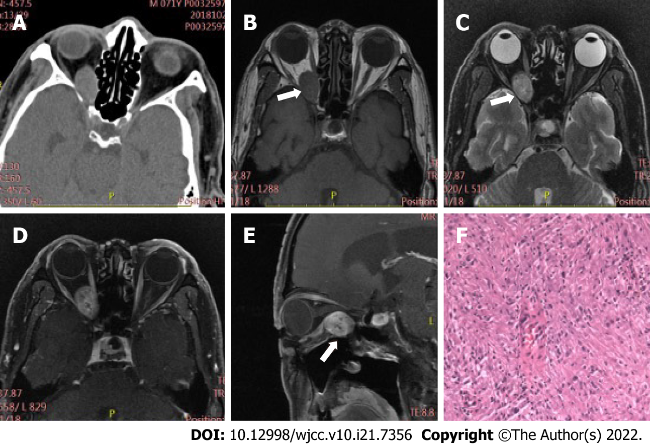
Figure 1 Case 1, 71-year-old male, right orbital schwannoma.
A: On plain computed tomography (CT) scan, oval soft tissue nodule with smooth edge can be seen in the right orbit (indicated by the arrow), the size is about 25 mm × 16 mm × 15 mm, and the CT value is approximately 42 Hu; B-E: Magnetic resonance imaging showed that the focus was located in the extrapyramidal space of the muscle below the right orbit (indicated by the arrow), having slightly long T1, T2 signals, and the enhanced scan displayed uneven persistence moderate or obvious enhancement; F: Spindle cell can be seen microscopically.
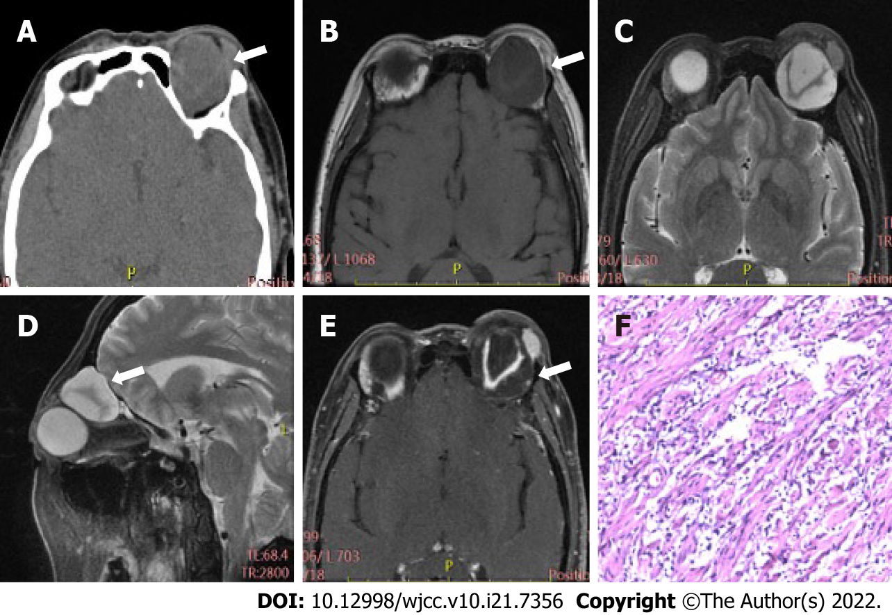
Figure 2 Case 2, 53-year-old female, left orbital myxoid neurofibroma.
A: A non-uniform density mass of left orbit with a size of 33 mm × 30 mm × 21 mm was displayed by computed tomography (CT) (as shown by the arrow); the CT value was 21-60 Hu; B-E: Magnetic resonance imaging showed that the focus, which was mainly cystic (long T1 and T2 signals), was located in the extrapyramidal space above the left orbit (as shown by the arrow); "V" shaped nerve fibers were observed (equal T1 and T2 signals); on contrast-enhanced scan, the cystic part was not enhanced, while the solid part showed continuous and obvious enhancement; F: Microscopically, there is a large amount of mucus around nerve fiber cells.
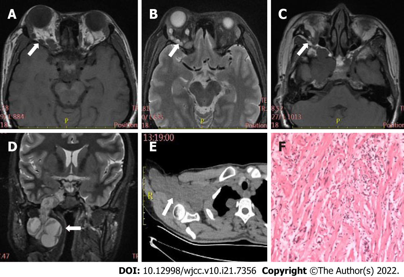
Figure 3 Case 3, 37-year-old female, plexiform neurofibroma.
A and B: Magnetic resonance imaging plain scan showed nodules, with a diameter of 4-5 mm, had multiple slightly long T1 and long/slightly long T2 signals in the right orbit (indicated by the arrow); C and D: The focus grew into the brain through the supraorbital and inferior fissure (indicated by the arrow). An irregular mass, which was mainly cystic (long T1, T2 signals), with "V" shaped nerve fibers (equal T1 and T2 signals), could be seen in the right parasellar, pterygopalatine as well as infratemporal fossa; E: Plain chest computed tomography (CT) scan indicated multiple irregular and slightly low-density nodules, together with masses, located in the right brachial plexus distribution and the subpleural region, having an average CT value of approximately 36 Hu; F: Microscopically, nerve fiber cells with mucus around them could be seen.
- Citation: Dai M, Wang T, Wang JM, Fang LP, Zhao Y, Thakur A, Wang D. Imaging characteristics of orbital peripheral nerve sheath tumors: Analysis of 34 cases. World J Clin Cases 2022; 10(21): 7356-7364
- URL: https://www.wjgnet.com/2307-8960/full/v10/i21/7356.htm
- DOI: https://dx.doi.org/10.12998/wjcc.v10.i21.7356


