Copyright
©The Author(s) 2022.
World J Clin Cases. Jan 14, 2022; 10(2): 741-746
Published online Jan 14, 2022. doi: 10.12998/wjcc.v10.i2.741
Published online Jan 14, 2022. doi: 10.12998/wjcc.v10.i2.741
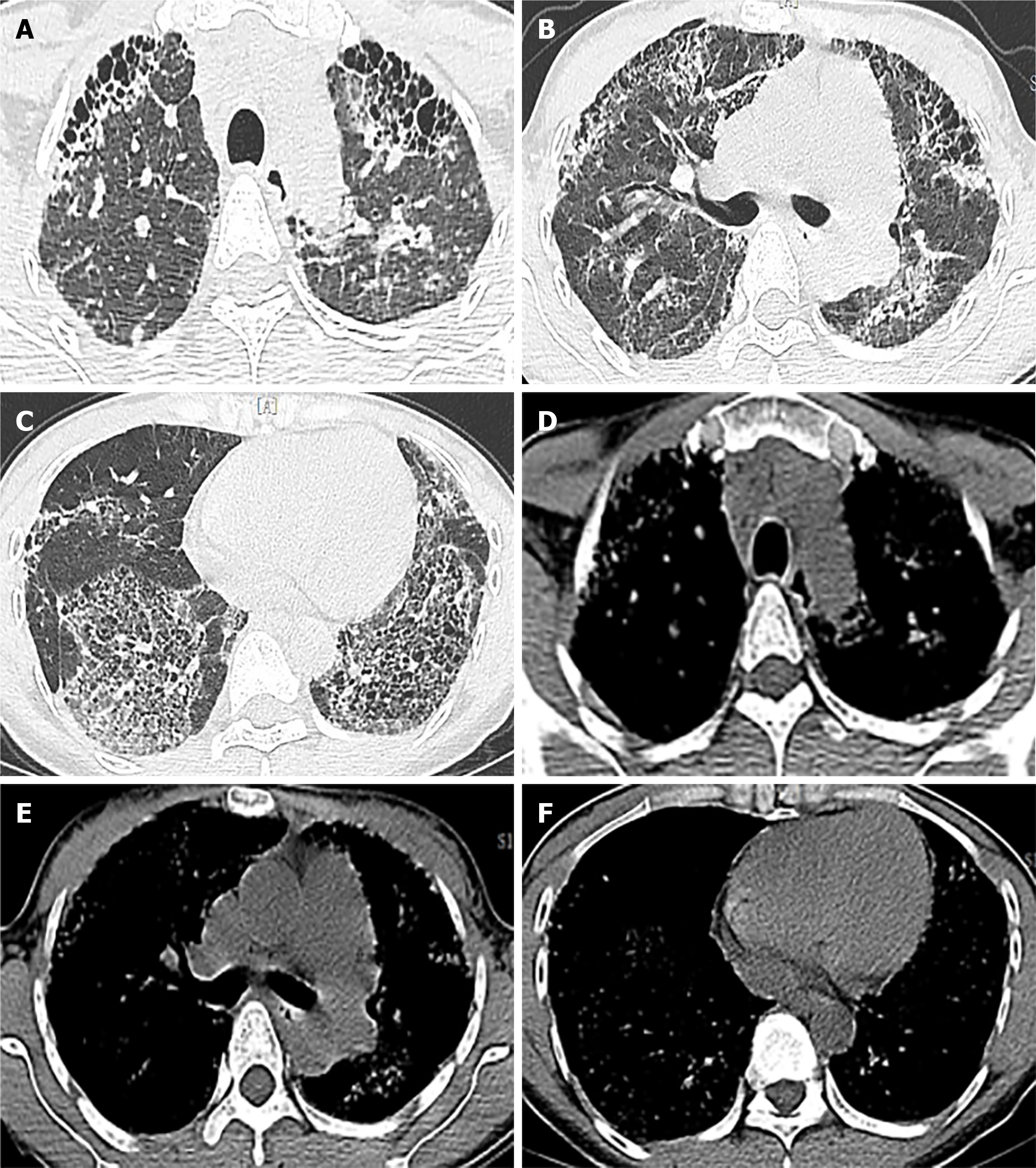
Figure 1 Chest high-resolution computed tomography (HRCT) images.
A-C: showing traction bronchiectasis and extensive fibrosis, reticular changes (especially in the outer lung band and the double lower lung); D-F: showing the corresponding mediastinal window.
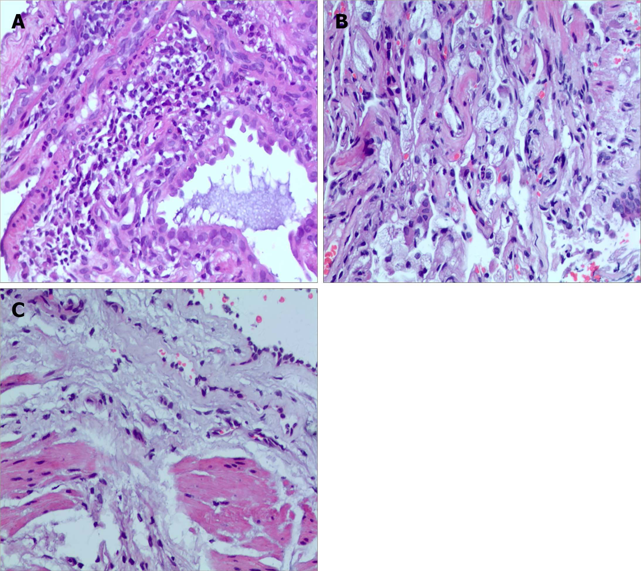
Figure 2 Pathological findings of transbronchial lung biopsy (TBLB) and computed tomography (CT)-guided percutaneous lung puncture, hematoxylin and eosin staining (×400).
A: chronic lymphocyte infiltration; B: foamy macrophages infiltration and diffuse, relatively uniform alveolar septal thickness; C: hyperplasia of fibrous tissue in the bronchial wall, resulting in fibrosis.
- Citation: Wang M, Fang HH, Jiang ZF, Ye W, Liu RY. Occupational fibrotic hypersensitivity pneumonia in a halogen dishes manufacturer: A case report. World J Clin Cases 2022; 10(2): 741-746
- URL: https://www.wjgnet.com/2307-8960/full/v10/i2/741.htm
- DOI: https://dx.doi.org/10.12998/wjcc.v10.i2.741


