Copyright
©The Author(s) 2022.
World J Clin Cases. Jan 14, 2022; 10(2): 709-716
Published online Jan 14, 2022. doi: 10.12998/wjcc.v10.i2.709
Published online Jan 14, 2022. doi: 10.12998/wjcc.v10.i2.709
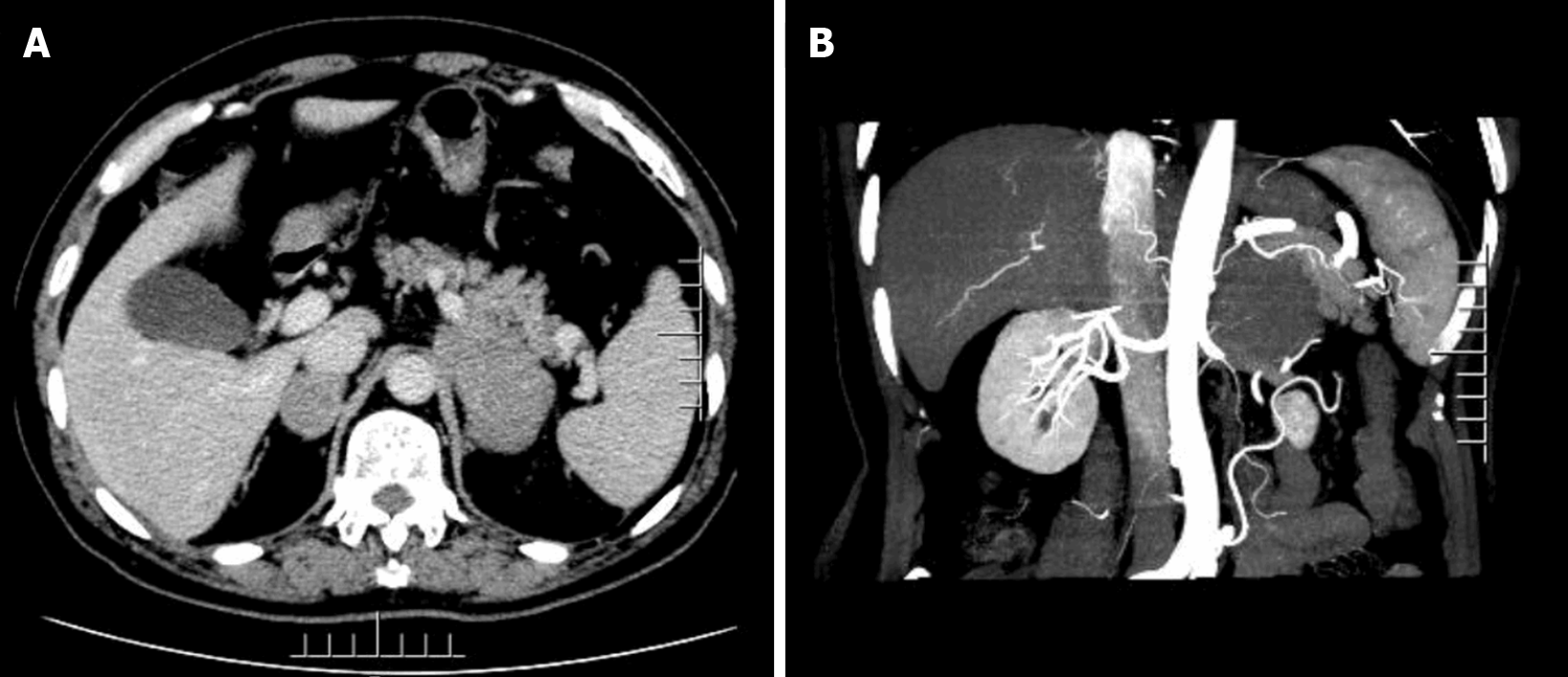
Figure 1 Preoperative abdominal enhanced computed tomography.
A: Irregular masses were observed in the bilateral adrenal glands, with the larger masses being located on the left side, with a size of approximately 8.0 cm × 4.3 cm. No obvious enhancement was noted in the enhanced arterial phase, whereas uneven enhancement was observed in the portal phase; B: The left mass was surrounded the left renal artery, compressing the upper pole of the left kidney and part of the left renal vein. The boundary was irregular, while no obvious enlarged lymph nodes were found behind the peritoneum.
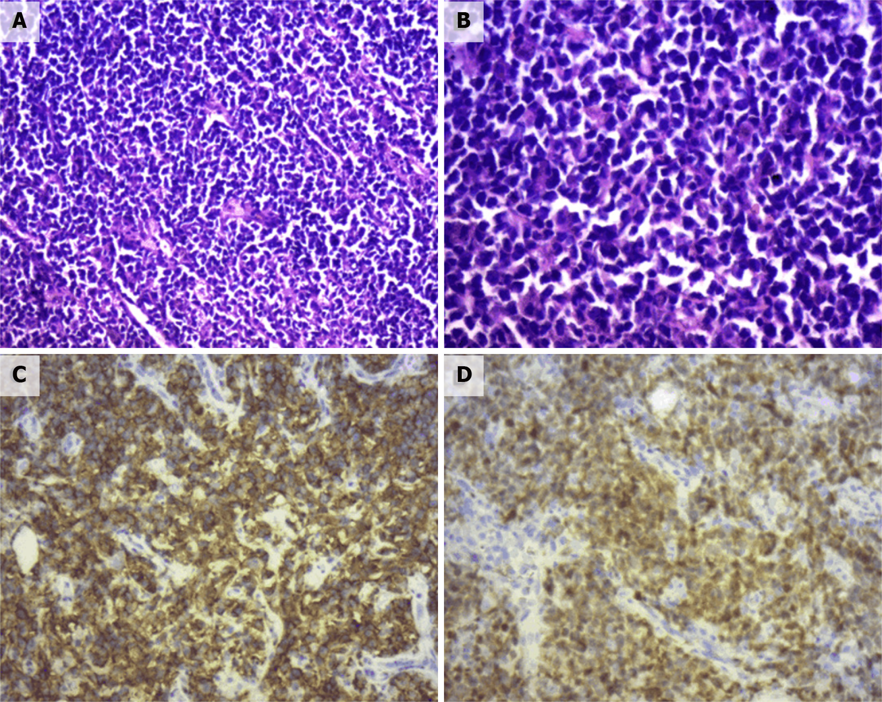
Figure 2 Pathological section.
A: Hematoxylin & eosin (HE, × 100). Diffuse infiltration and growth of tumor cells; B: HE (× 200). Tumor cells were composed of medium to large lymphoid cells, most of which were round and oval, double chromotropic or basophilic, containing less cytoplasm and large nuclei; C: Immunohistochemical SP method of CD20 staining. Strong positively stained cell membrane in all tumor cells (the brownish yellow part of the picture); D: Immunohistochemical SP method of CD20 staining. Strong positively stained cell membrane in all tumor cells (the brownish yellow part of the picture).
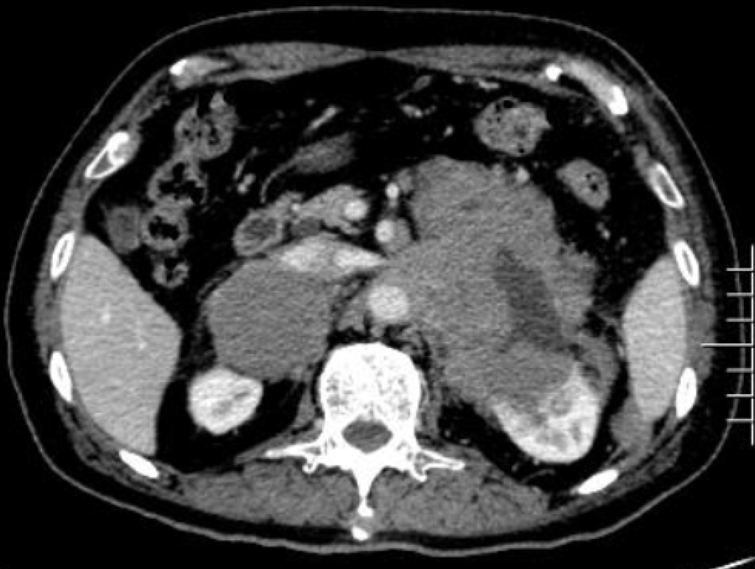
Figure 3 Re-examination of abdominal enhanced computed tomography 1 mo after surgery.
Multiple soft tissue opacity in bilateral retroperitoneum, uneven enhancement, compression in the liver, spleen and pancreas, unclear boundary, unclear adrenal glands on the right side, and a soft tissue mass protruding into the kidney on the left retroperitoneum.
- Citation: Fan ZN, Shi HJ, Xiong BB, Zhang JS, Wang HF, Wang JS. Primary adrenal diffuse large B-cell lymphoma with normal adrenal cortex function: A case report. World J Clin Cases 2022; 10(2): 709-716
- URL: https://www.wjgnet.com/2307-8960/full/v10/i2/709.htm
- DOI: https://dx.doi.org/10.12998/wjcc.v10.i2.709


