Copyright
©The Author(s) 2022.
World J Clin Cases. Jul 6, 2022; 10(19): 6702-6709
Published online Jul 6, 2022. doi: 10.12998/wjcc.v10.i19.6702
Published online Jul 6, 2022. doi: 10.12998/wjcc.v10.i19.6702
Figure 1 Radical resection of the anterior abdominal wall tumor was performed.
A: A mass located on the lower abdomen with infection and the surface was covered with a white crust; B: The large defect after radical resection of the anterior abdominal wall tumor; C: The reconstructed abdominal wall with a Bard composite mesh and the local flap designed to help close the external covering of abdomen; D: Appearance of the abdominal reconstruction after surgery immediately.
Figure 2 Computed tomography images.
A: A pelvic magnetic resonance imaging showed a huge mass located on the middle of the anterior abdominal wall; B: The computed tomography showed the mass on the anterior abdominal wall invading the bladder.
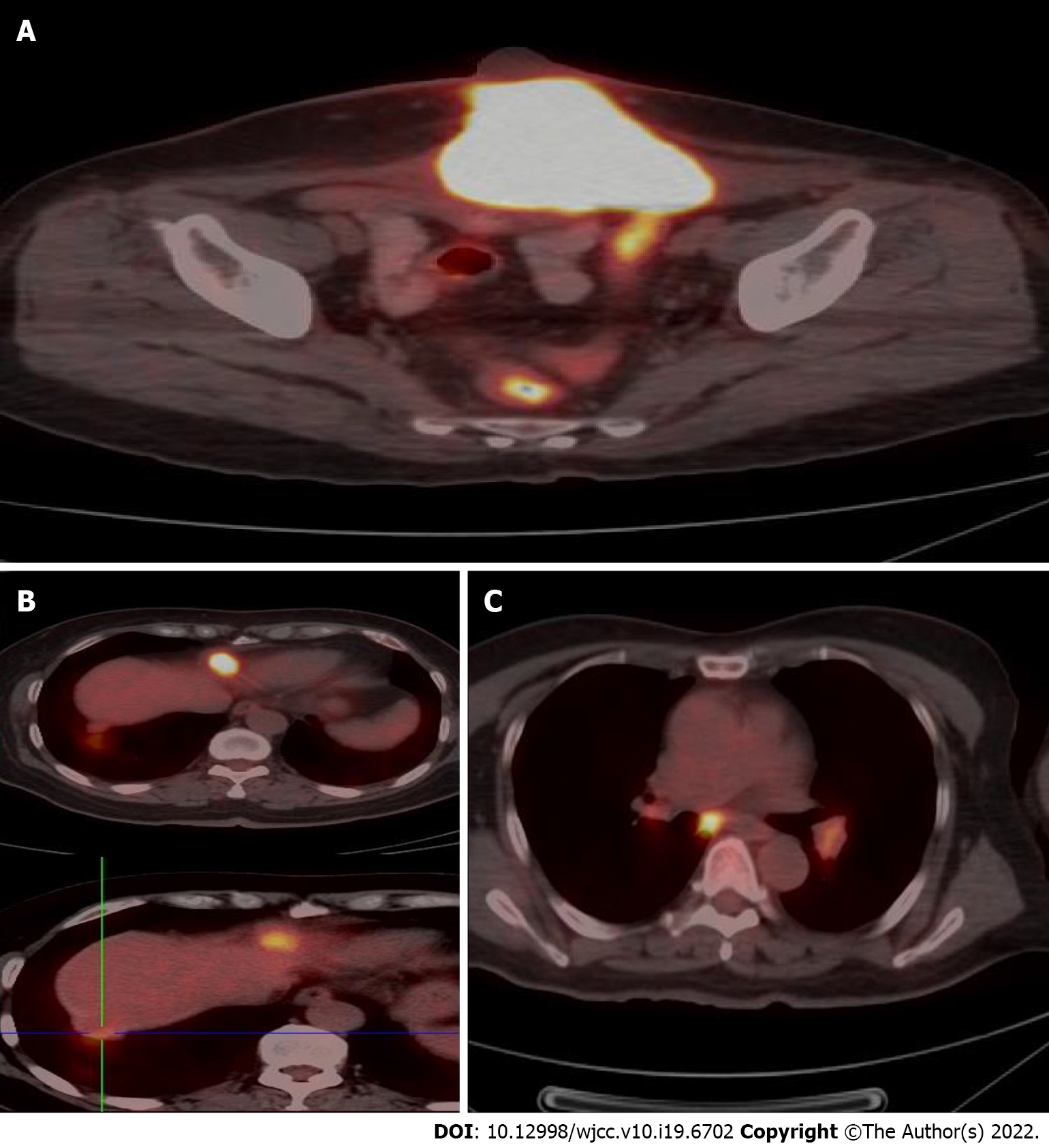
Figure 3 Positron emission tomography-computed tomography examination.
A: Increased fluorodeoxyglucose (FDG) uptake in the mass on the abdominal wall, with an SUVmax of 21.5 on the fusion images; B: Increased FDG uptake in two nodules under the liver capsule, with an SUVmax of 10.9 and 3.9 respectively on the fusion images; C: Increased FDG uptake in mediastinum and hilum lymph nodes with an SUVmax of 3.5-6.0 on the fusion images.
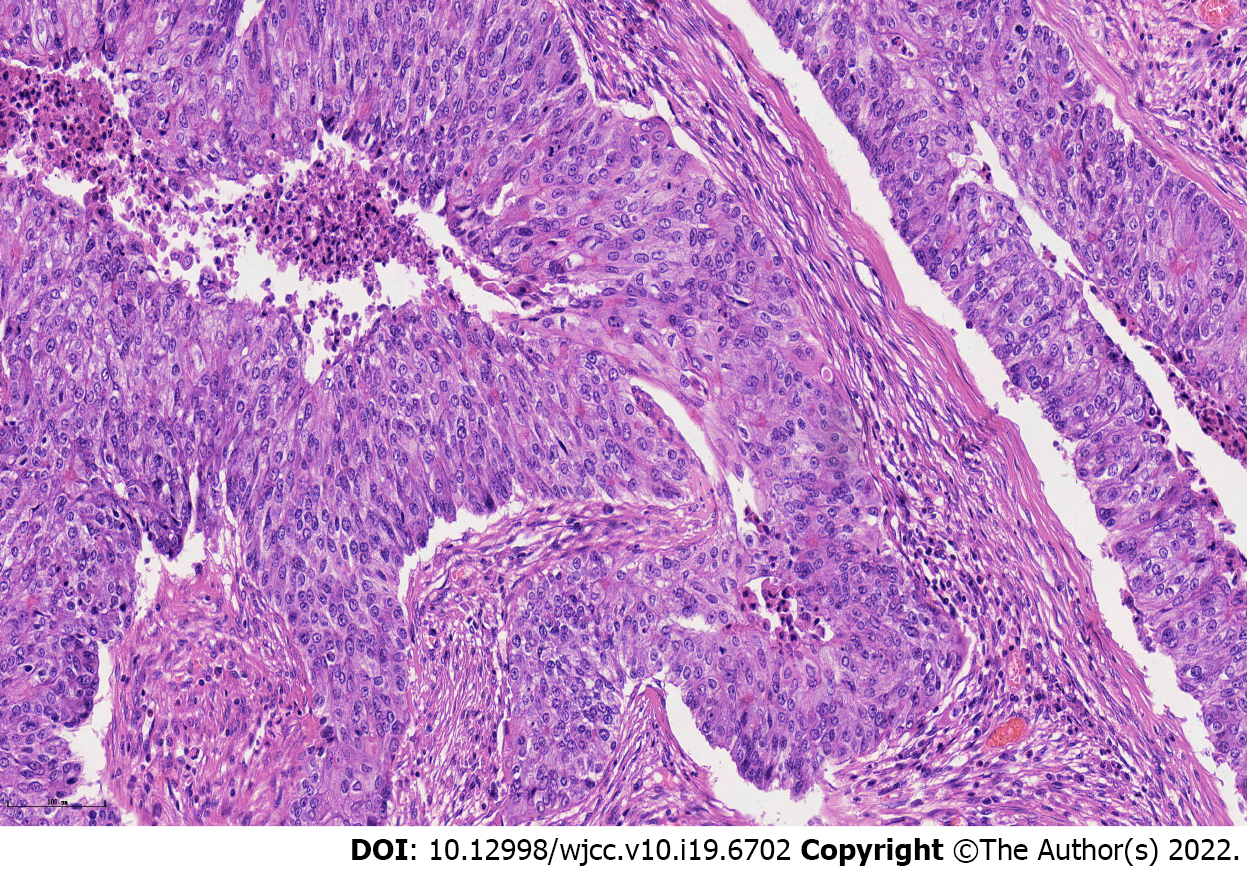
Figure 4 Hemotoxylin and eosin staining of endometrioid carcinoma, × 200 magnification.
- Citation: Wang JY, Wang ZQ, Liang SC, Li GX, Shi JL, Wang JL. Plastic surgery for giant metastatic endometrioid adenocarcinoma in the abdominal wall: A case report and review of literature. World J Clin Cases 2022; 10(19): 6702-6709
- URL: https://www.wjgnet.com/2307-8960/full/v10/i19/6702.htm
- DOI: https://dx.doi.org/10.12998/wjcc.v10.i19.6702


