Copyright
©The Author(s) 2022.
World J Clin Cases. Jul 6, 2022; 10(19): 6543-6547
Published online Jul 6, 2022. doi: 10.12998/wjcc.v10.i19.6543
Published online Jul 6, 2022. doi: 10.12998/wjcc.v10.i19.6543
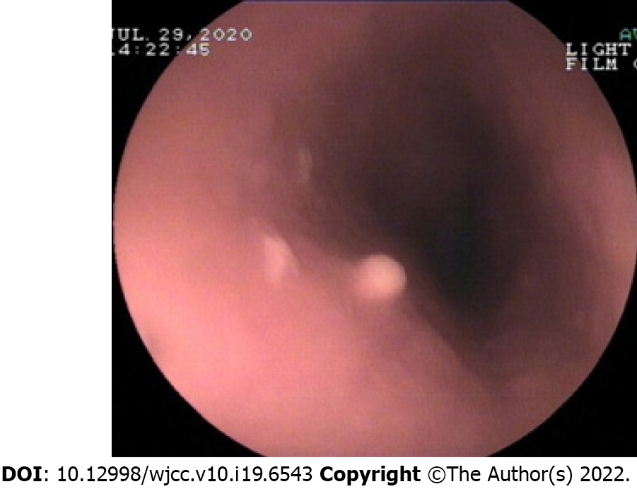
Figure 1 Endoscopy of esophageal granular cell tumor.
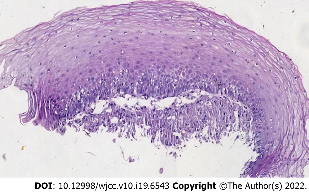
Figure 2 HE staining X200.
The tumor cells were closely arranged in a cordlike pattern,
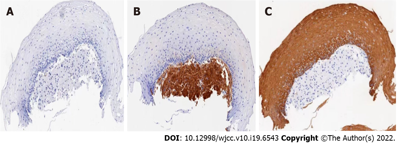
Figure 3 Immunohistochemical S100 positive.
A: Contrast diagram not stained with S100; B and C: the nucleus and cytoplasm of granulosa cell tumor are brown and yellow by S-100 staining.
- Citation: Chen YL, Zhou J, Yu HL. Esophageal granular cell tumor: A case report. World J Clin Cases 2022; 10(19): 6543-6547
- URL: https://www.wjgnet.com/2307-8960/full/v10/i19/6543.htm
- DOI: https://dx.doi.org/10.12998/wjcc.v10.i19.6543


