Copyright
©The Author(s) 2022.
World J Clin Cases. Jul 6, 2022; 10(19): 6446-6455
Published online Jul 6, 2022. doi: 10.12998/wjcc.v10.i19.6446
Published online Jul 6, 2022. doi: 10.12998/wjcc.v10.i19.6446
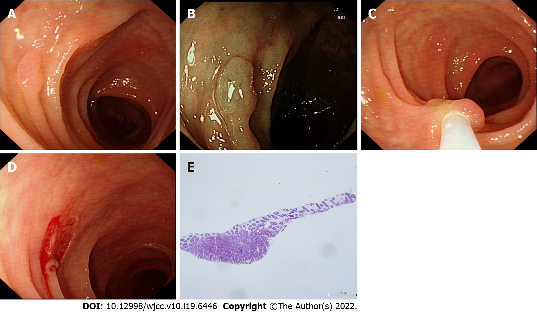
Figure 1 The process of cold snare polypectomy.
A: Polyp approximately 0.7 cm in diameter identified under the white light of colonoscopy; B: Polyp observed under narrow band imaging; C: Tightening of the snare during cold snare polypectomy (CSP); D: Wound after CSP; E: Postoperative pathological tissue specimen.
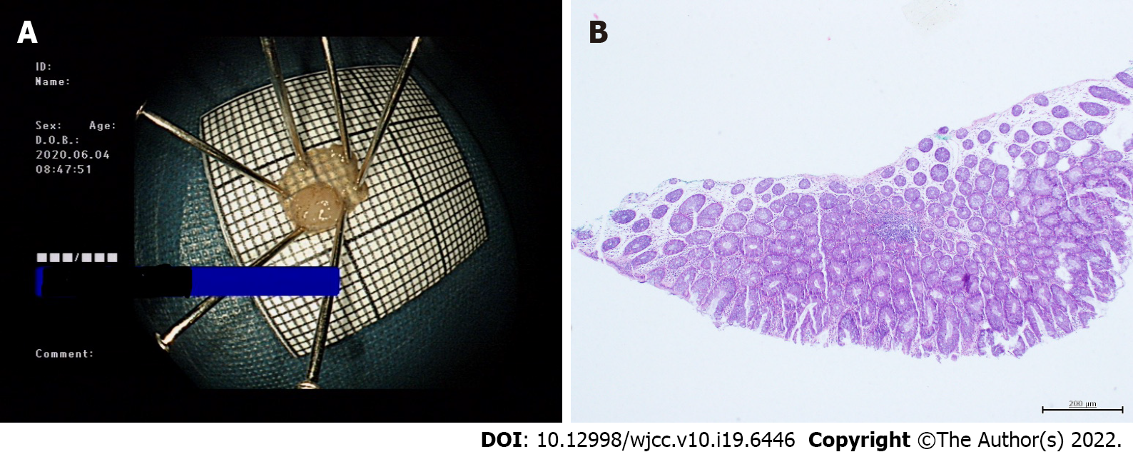
Figure 2 Tissue specimen of cold snare polypectomy.
A: Gross specimen of cold snare polypectomy (CSP); B: Cutting edge of CSP under high magnification.
- Citation: Meng QQ, Rao M, Gao PJ. Effect of cold snare polypectomy for small colorectal polyps. World J Clin Cases 2022; 10(19): 6446-6455
- URL: https://www.wjgnet.com/2307-8960/full/v10/i19/6446.htm
- DOI: https://dx.doi.org/10.12998/wjcc.v10.i19.6446


