Copyright
©The Author(s) 2022.
World J Clin Cases. Jun 6, 2022; 10(16): 5456-5462
Published online Jun 6, 2022. doi: 10.12998/wjcc.v10.i16.5456
Published online Jun 6, 2022. doi: 10.12998/wjcc.v10.i16.5456
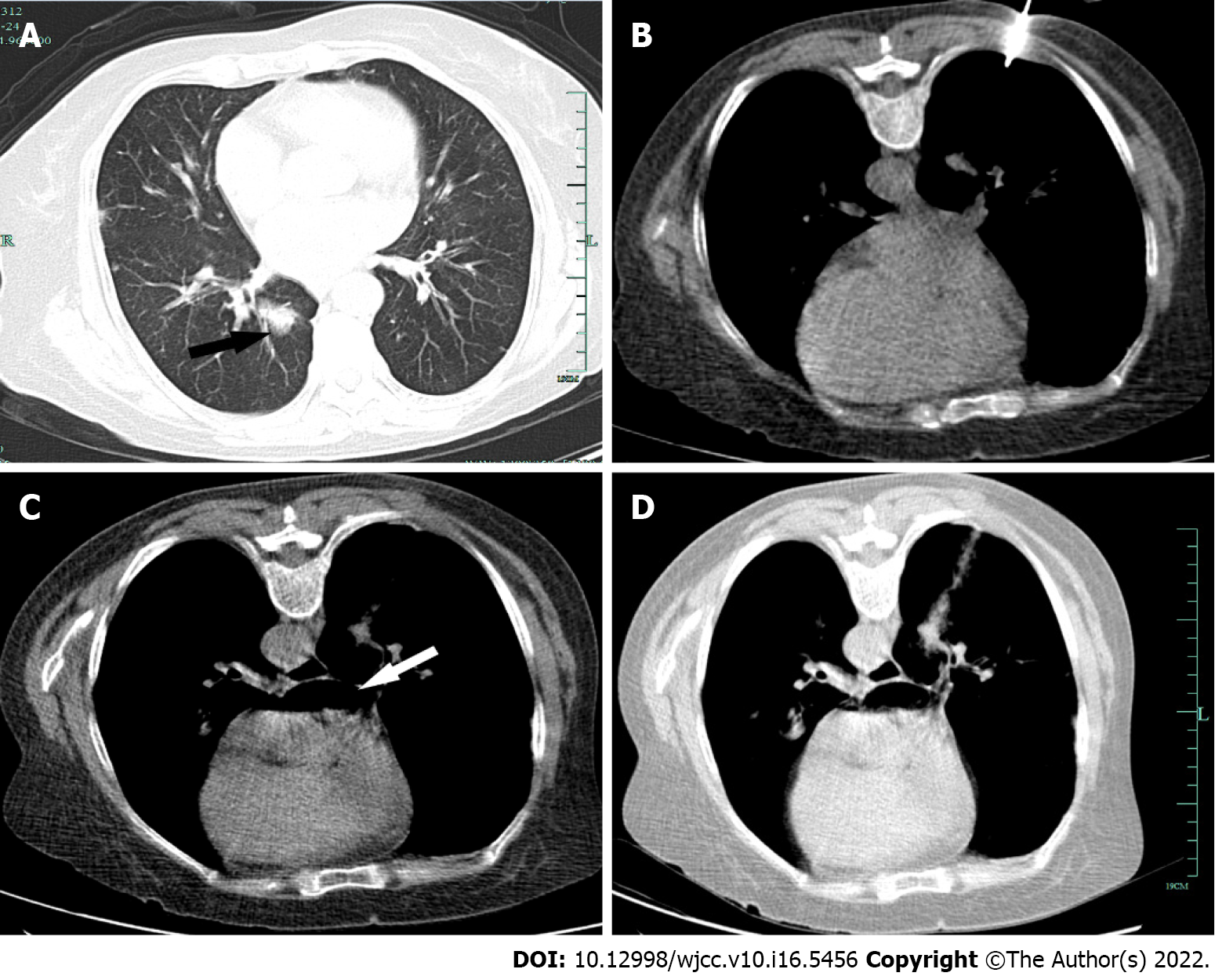
Figure 1 Computed tomography findings in the patient before and after biopsy.
A: Prior to the procedure, computed tomography (CT) scanning was conducted to establish an appropriate needle trajectory, with the nodule of interest being located just on the posterior basal segment of the right lower lobe (black arrow); B: The co-axial 15-gauge needle of a core biopsy instrument was inserted through the nodule. During the first round of biopsy procedure, there was no air in the left atrium; C: After the second round of biopsy, a massive volume of air traveled to the left atrium (white arrow); D: CT scanning revealed a faint connection between blood vessels and the airways.
- Citation: Li YW, Chen C, Xu Y, Weng QP, Qian SX. Fatal left atrial air embolism as a complication of percutaneous transthoracic lung biopsy: A case report. World J Clin Cases 2022; 10(16): 5456-5462
- URL: https://www.wjgnet.com/2307-8960/full/v10/i16/5456.htm
- DOI: https://dx.doi.org/10.12998/wjcc.v10.i16.5456


