Copyright
©The Author(s) 2022.
World J Clin Cases. Jun 6, 2022; 10(16): 5435-5440
Published online Jun 6, 2022. doi: 10.12998/wjcc.v10.i16.5435
Published online Jun 6, 2022. doi: 10.12998/wjcc.v10.i16.5435
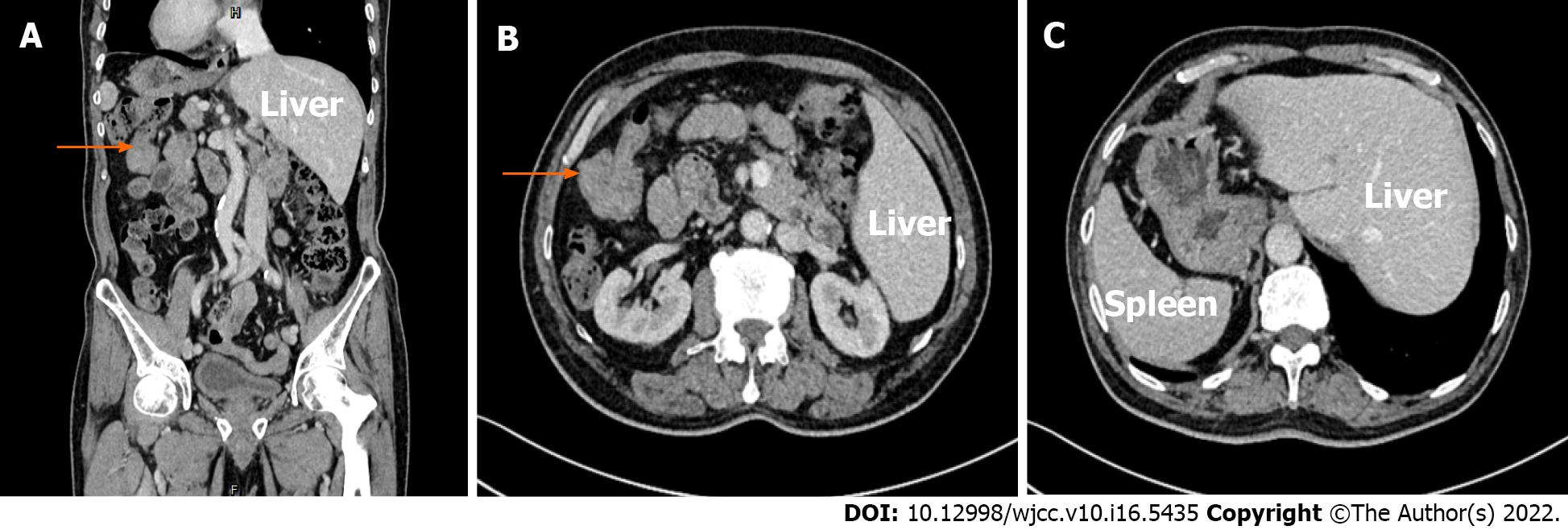
Figure 1 Computed tomography imaging.
A: Coronal plane of contrasted computed tomography (CT); B:The arrow indicates splenic flexure cancer; C: Transverse section of contrasted CT.
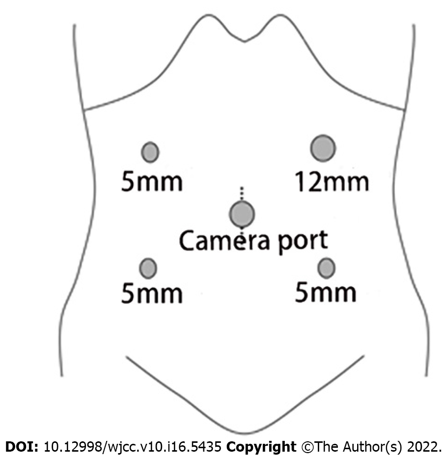
Figure 2 Trocar position.
The position of the trocar was in a mirror image arrangement. The 12-mm trocar for the operating surgeon was located in the left upper quadrant.
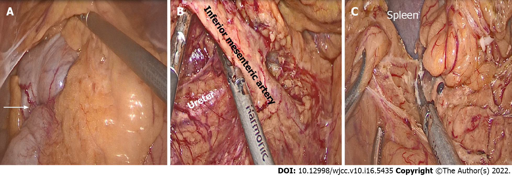
Figure 3 Intraoperative findings.
A: The arrow shows the position of the tumor; B: The inferior mesenteric artery was mobilized, and the right ureter was confirmed on the dorsal side; C: Mobilization of the colonic splenic flexure.
- Citation: Zheng ZL, Zhang SR, Sun H, Tang MC, Shang JK. Laparoscopic radical resection for situs inversus totalis with colonic splenic flexure carcinoma: A case report. World J Clin Cases 2022; 10(16): 5435-5440
- URL: https://www.wjgnet.com/2307-8960/full/v10/i16/5435.htm
- DOI: https://dx.doi.org/10.12998/wjcc.v10.i16.5435


