Copyright
©The Author(s) 2022.
World J Clin Cases. Jun 6, 2022; 10(16): 5428-5434
Published online Jun 6, 2022. doi: 10.12998/wjcc.v10.i16.5428
Published online Jun 6, 2022. doi: 10.12998/wjcc.v10.i16.5428
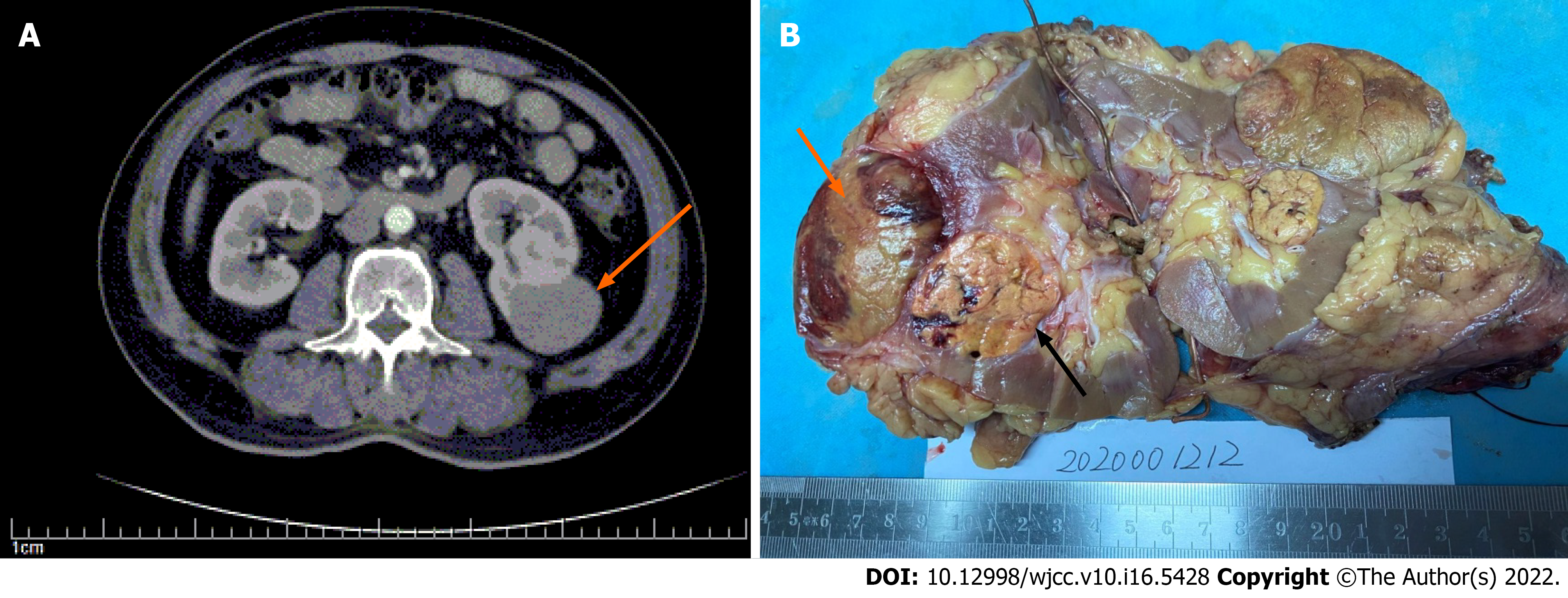
Figure 1 Macroscopic features and conventional computed tomography findings.
A: Conventional computed tomography demonstrated a single endophytic mass in the lower pole of the kidney, with contrast enhancement (orange arrow); B: Gross surface showed two lesions in the middle-lower pole of the kidney, papillary renal cell carcinoma (black arrow) and clear cell renal cell carcinoma (orange arrow).
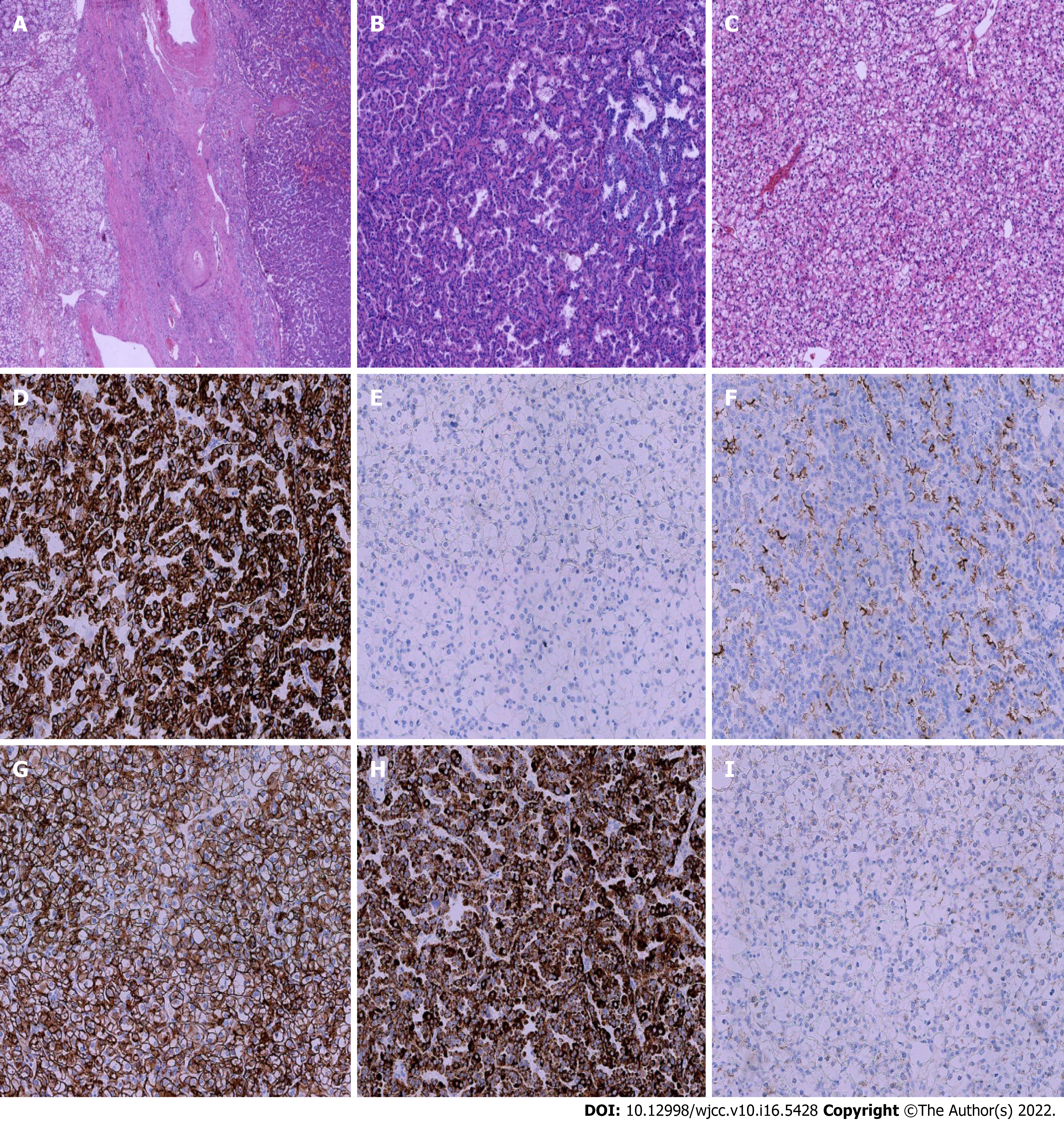
Figure 2 Histopathological findings.
A: Hematoxylin–eosin (HE) staining showed a clear border between clear cell renal cell carcinoma (CCRCC) (left) and papillary cell renal cell carcinoma (PRCC) (right) 200 ×; B: HE staining of PRCC 200 ×; C: HE staining of CCRCC 200 ×; D-E: Immunohistochemical (IHC) staining showed that CK7 was positive in PRCC and negative in CCRCC 200 ×; F-G: IHC staining showed that CD10 was diffusely positive in PRCC and in CCRCC 200 ×; H-I: IHC staining showed that P504s was positive in PRCC and negative in CCRCC.
- Citation: Yin J, Zheng M. Ipsilateral synchronous papillary and clear renal cell carcinoma: A case report and review of literature. World J Clin Cases 2022; 10(16): 5428-5434
- URL: https://www.wjgnet.com/2307-8960/full/v10/i16/5428.htm
- DOI: https://dx.doi.org/10.12998/wjcc.v10.i16.5428


