Copyright
©The Author(s) 2022.
World J Clin Cases. Jun 6, 2022; 10(16): 5387-5393
Published online Jun 6, 2022. doi: 10.12998/wjcc.v10.i16.5387
Published online Jun 6, 2022. doi: 10.12998/wjcc.v10.i16.5387
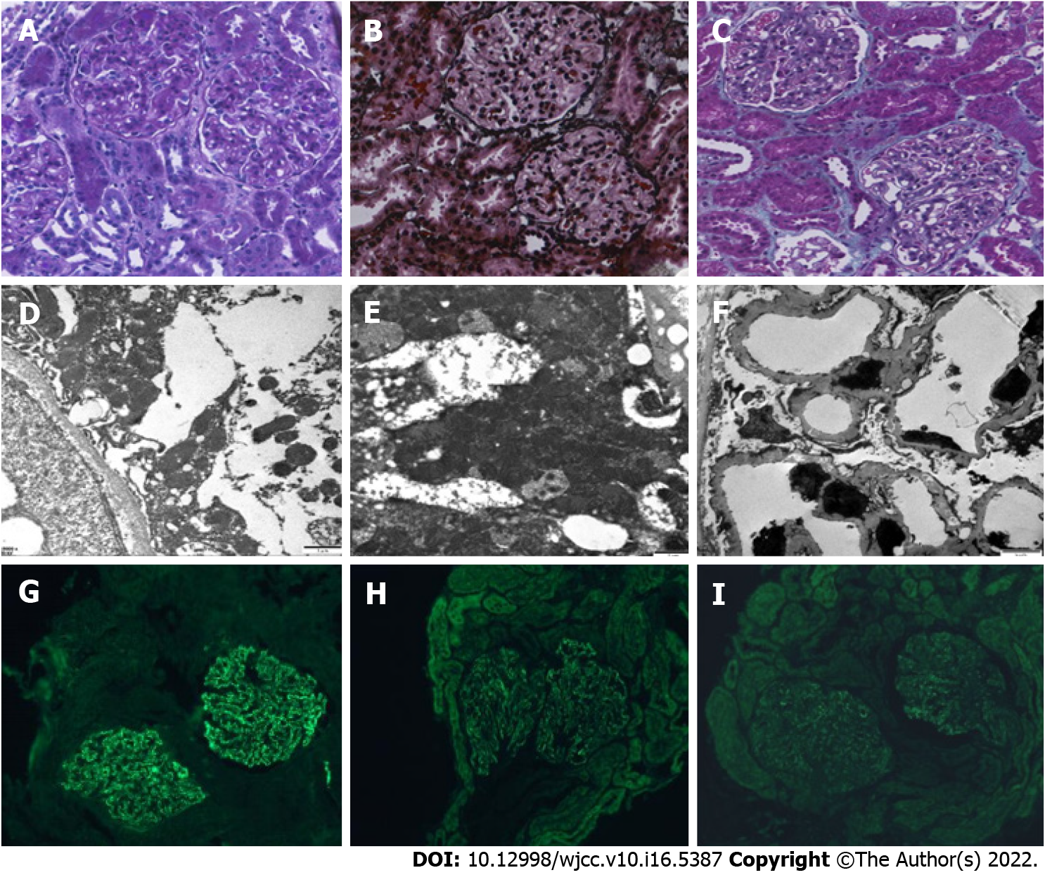
Figure 1 Representative images of pathological changes on renal biopsy.
Diffuse thickening of the basement membrane with several spiky formations, subepithelial deposition of fuchsinophilic protein, and vacuolar and granular degeneration of renal tubular epithelial cells were observed. A: Light microscopy (Periodic acid-Schiff, × 400); B: Light microscopy (Periodic acid-silver methenamine, × 400); C: Light microscopy (Masson staining, × 400); D-F: Irregular thickening of the basement membrane, electron-dense deposits in the subepithelial and intrabasal areas, and diffuse fusion of the foot processes (electron microscopy, × 6000); G: Depositions of immunoglobulin G (IgG) along the mesangial area and the capillary wall (immunohistochemical staining, × 400); H: Depositions of IgG1 along the mesangial area and the capillary wall (immunohistochemical staining, × 400); I: Depositions of IgG4 along the mesangial area and the capillary wall. Positivity for phospholipase A2 receptor and negativity for thrombospondin type-1 domain-containing 7A are shown (immunohistochemical staining, × 400).
- Citation: Cui KH, Zhang H, Tao YH. Idiopathic membranous nephropathy in children: A case report. World J Clin Cases 2022; 10(16): 5387-5393
- URL: https://www.wjgnet.com/2307-8960/full/v10/i16/5387.htm
- DOI: https://dx.doi.org/10.12998/wjcc.v10.i16.5387


