Copyright
©The Author(s) 2022.
World J Clin Cases. Jun 6, 2022; 10(16): 5380-5386
Published online Jun 6, 2022. doi: 10.12998/wjcc.v10.i16.5380
Published online Jun 6, 2022. doi: 10.12998/wjcc.v10.i16.5380
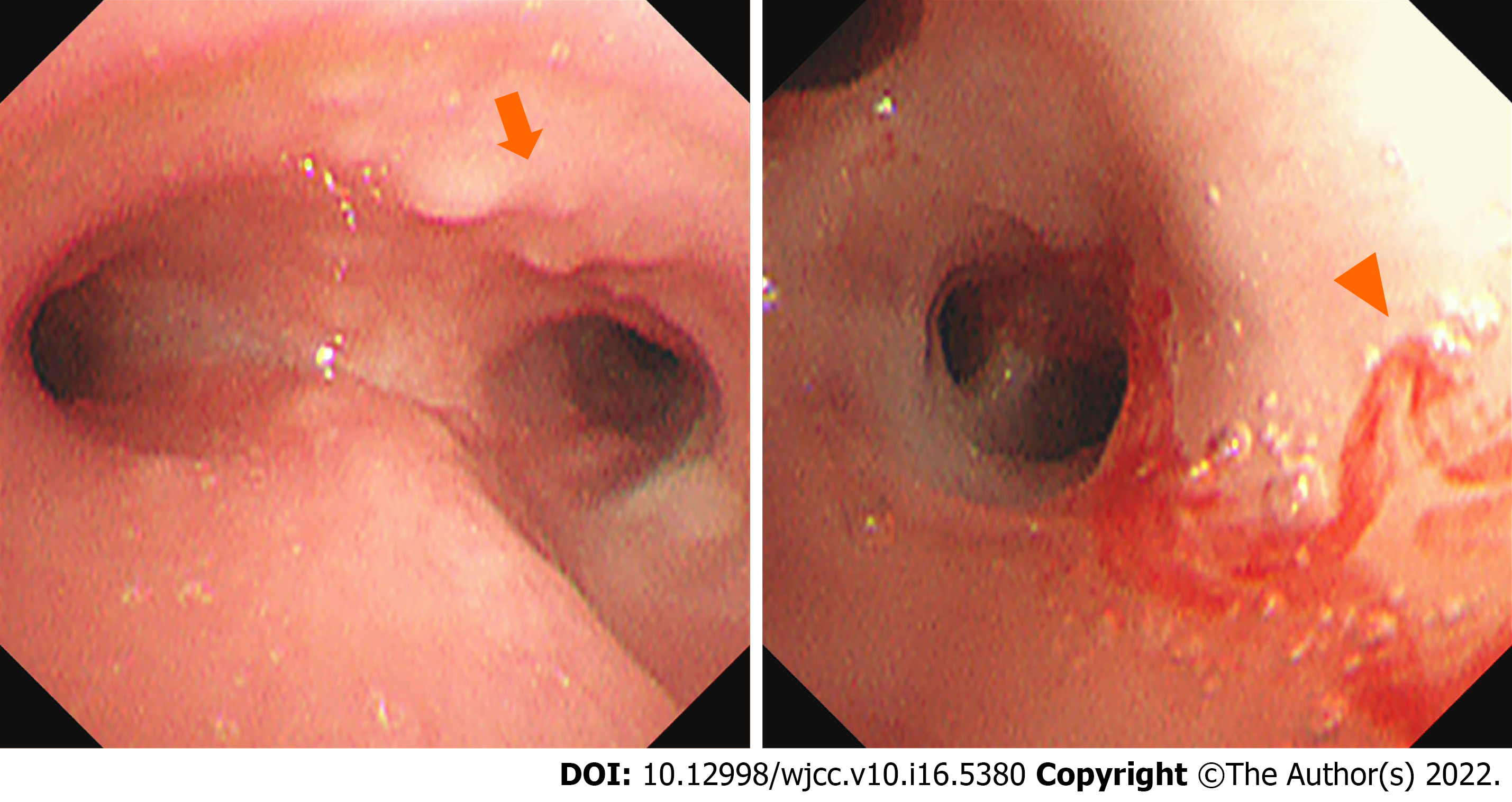
Figure 1 White nodules in trachea and the left main bronchus are observed (arrow).
The mucosa of the posterior basal segment of the left lower lobe is pale, and mild hemorrhage in the posterior basal segment of the left lower lobe (arrowhead) is observed.
Figure 2 Lymphoproliferative lesions with necrosis.
A: The proliferated and infiltrated lymphocytes are moderately large, the nucleus is round, and some nucleoli can be observed (HE × 100); B: Cells infiltrating blood vessels can be observed (HE × 200); C: Small lymphocytes, plasma cells, and histiocytes are observed in the background, (HE × 400).
Figure 3 Chest computed tomography.
A: Multiple nodules and mass shadows (arrows) are observed in the lower lobe of the right and left lungs, and significant bilateral lymphadenopathy in the hilum and mediastinum is visible; B: Multiple nodules and mass shadows are almost unchanged in the lower lobe of the right and left lungs (arrowhead) after anti-infective treatment. Bilateral hilar, mediastinal, and axillary lymphadenopathy is noted.
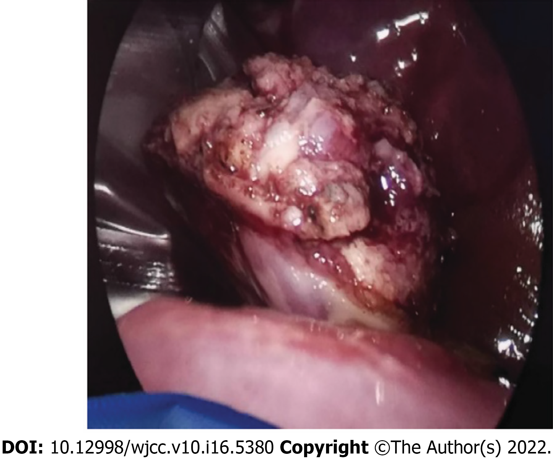
Figure 4 Thoracoscopic wedge resection of left lower lung.
A small amount of yellowish effusion, pleural thickening, fascicular adhesion between the lower lobe of the left lung and pleura, and a hard mass with unclear boundaries in the basal segment of the lower lobe of the left lung are observed in the pleural cavity.
- Citation: Yao JW, Qiu L, Liang P, Liu HM, Chen LN. Pulmonary lymphomatoid granulomatosis in a 4-year-old girl: A case report. World J Clin Cases 2022; 10(16): 5380-5386
- URL: https://www.wjgnet.com/2307-8960/full/v10/i16/5380.htm
- DOI: https://dx.doi.org/10.12998/wjcc.v10.i16.5380


