Copyright
©The Author(s) 2022.
World J Clin Cases. Jun 6, 2022; 10(16): 5359-5364
Published online Jun 6, 2022. doi: 10.12998/wjcc.v10.i16.5359
Published online Jun 6, 2022. doi: 10.12998/wjcc.v10.i16.5359
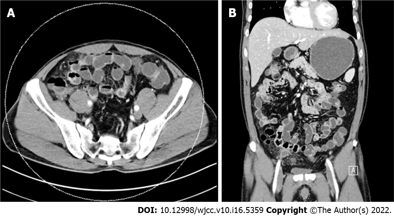
Figure 1 Abdominal computed tomography.
There are diffuse omental infiltrates and some peritoneal thickening with small lymph nodes in common hepatic, hepaticoduodenal, small bowel mesenteric, and para-aortic areas. A: Axial view; B: Coronal view.
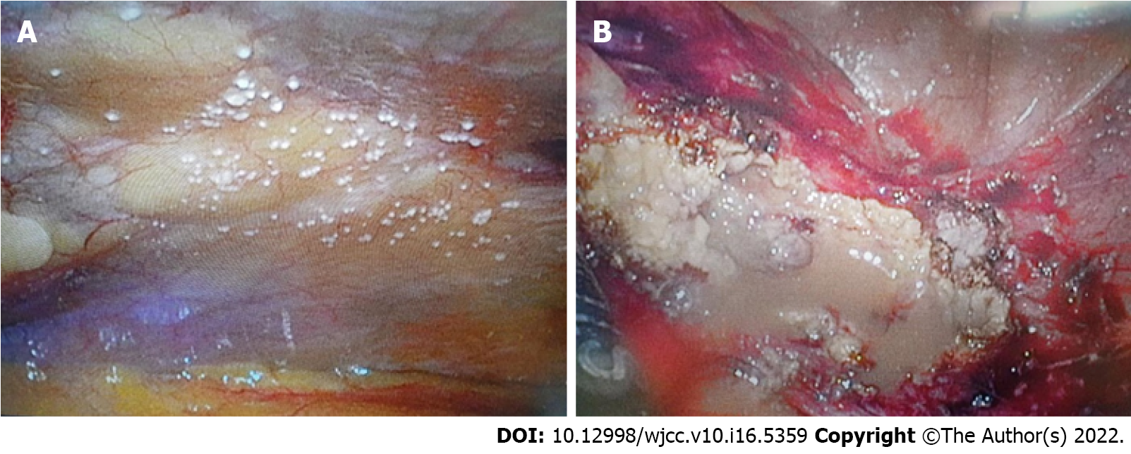
Figure 2 Laparoscopic findings.
A: Peritoneal nodules mimicked tuberculous nodules; B: Abscess pocket of the omentum is noted in the right pelvic wall around the anteroiliac area.
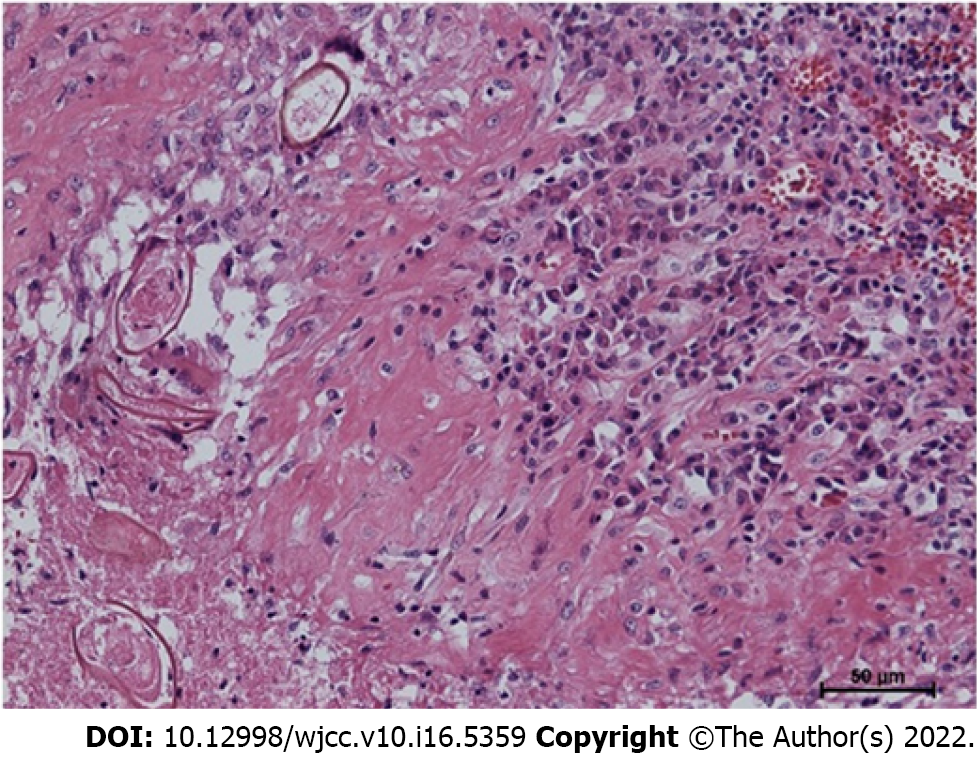
Figure 3 Pathological finding.
The parasite eggs are shown (hematoxylin and eosin stain, × 50).
- Citation: Choi JW, Lee CM, Kim SJ, Hah SI, Kwak JY, Cho HC, Ha CY, Jung WT, Lee OJ. Ectopic peritoneal paragonimiasis mimicking tuberculous peritonitis: A care report. World J Clin Cases 2022; 10(16): 5359-5364
- URL: https://www.wjgnet.com/2307-8960/full/v10/i16/5359.htm
- DOI: https://dx.doi.org/10.12998/wjcc.v10.i16.5359


