Copyright
©The Author(s) 2022.
World J Clin Cases. May 26, 2022; 10(15): 5111-5118
Published online May 26, 2022. doi: 10.12998/wjcc.v10.i15.5111
Published online May 26, 2022. doi: 10.12998/wjcc.v10.i15.5111
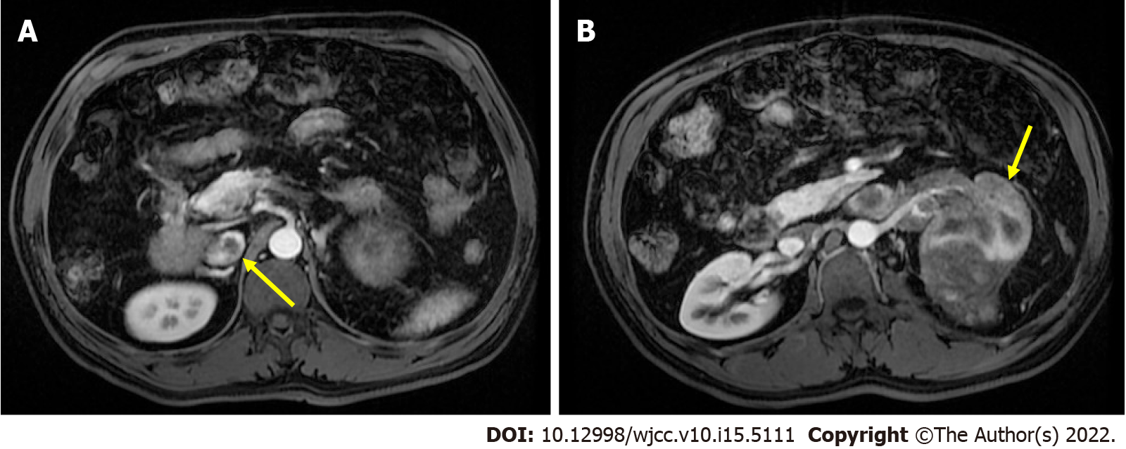
Figure 1 Abdominal magnetic resonance imaging for preoperative evaluation.
A: The inferior vena cava (IVC) tumor thrombus was at intrahepatic level, which was graded as level II (extends into the IVC, > 2 cm above the renal vein but below the hepatic veins; indicated by yellow arrow); B: Left renal tumor with renal vein invasion (indicated by yellow arrow).
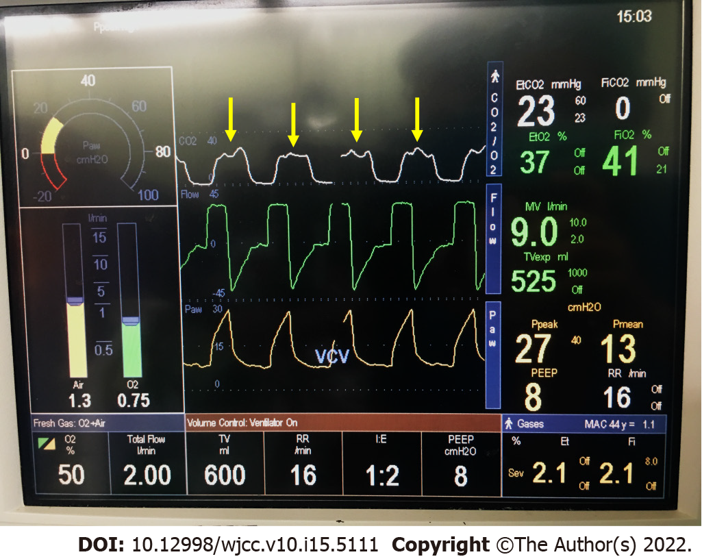
Figure 2 Intraoperative capnography when acute pulmonary embolism was suspected.
Oscillating of the plateau phase (phase III, indicated by yellow arrow) of capnography was observed in each breath.
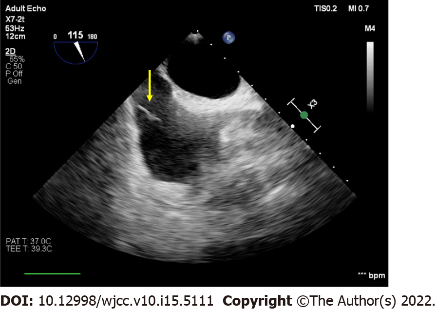
Figure 3 Intraoperative transesophageal echocardiography.
Middle-esophageal bicaval view of transesophageal echocardiography showed tumor thrombus in inferior vena cava (indicated by yellow arrow).
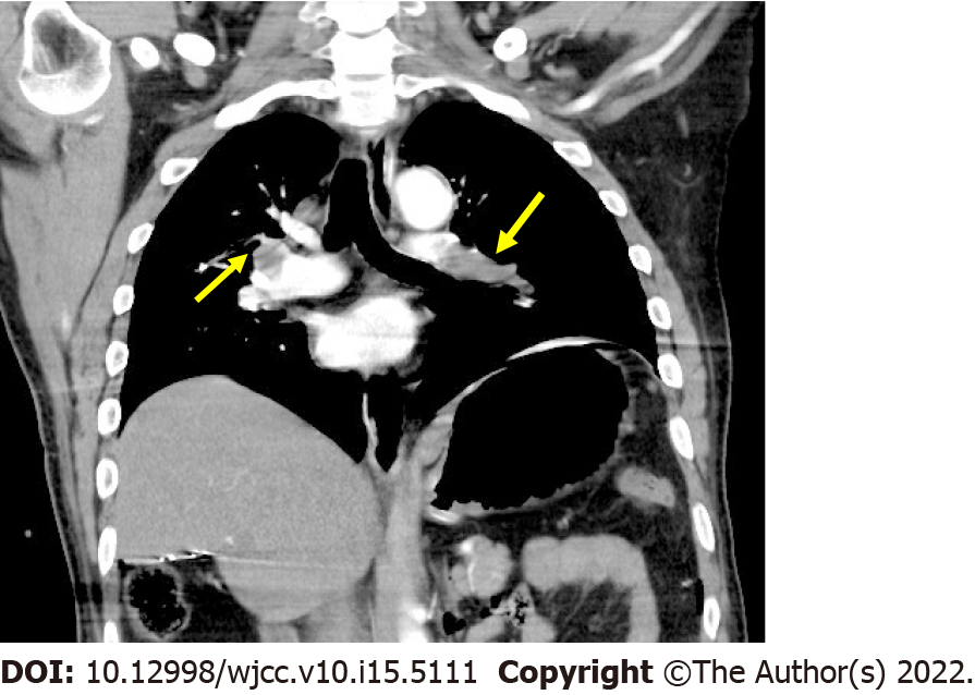
Figure 4 Postoperative chest computer tomography pulmonary angiogram.
Filling defects was observed in bilateral pulmonary arteries (indicated by yellow arrow).
- Citation: Hsu PY, Wu EB. Anesthetic management for intraoperative acute pulmonary embolism during inferior vena cava tumor thrombus surgery: A case report. World J Clin Cases 2022; 10(15): 5111-5118
- URL: https://www.wjgnet.com/2307-8960/full/v10/i15/5111.htm
- DOI: https://dx.doi.org/10.12998/wjcc.v10.i15.5111


