Copyright
©The Author(s) 2022.
World J Clin Cases. May 26, 2022; 10(15): 5088-5096
Published online May 26, 2022. doi: 10.12998/wjcc.v10.i15.5088
Published online May 26, 2022. doi: 10.12998/wjcc.v10.i15.5088
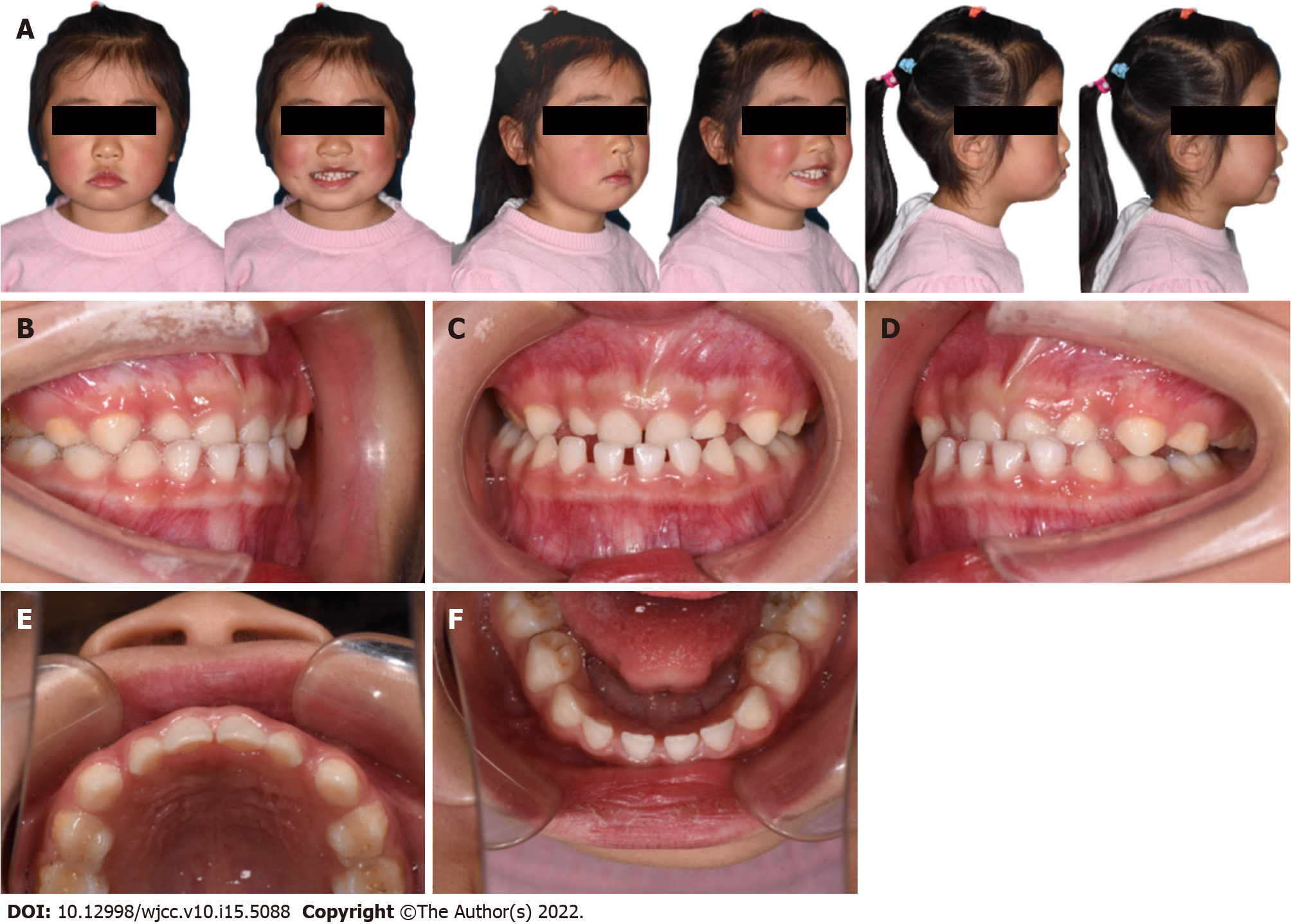
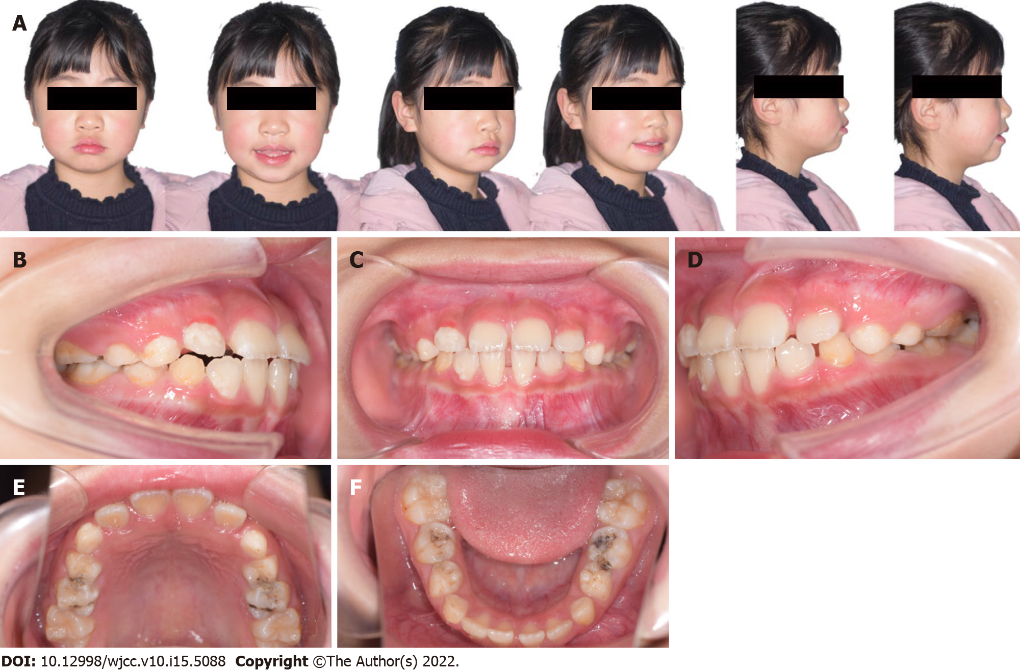
Figure 1 Pre-treatment facial and intra-oral photographs.
A: Pre-treatment facial photos showing presence of facial asymmetry; B: Lateral view of right side; C: Frontal view; D: Lateral view of left side; E: Occlusal view of the maxillary arch; F: Occlusal view of the mandibular arch.
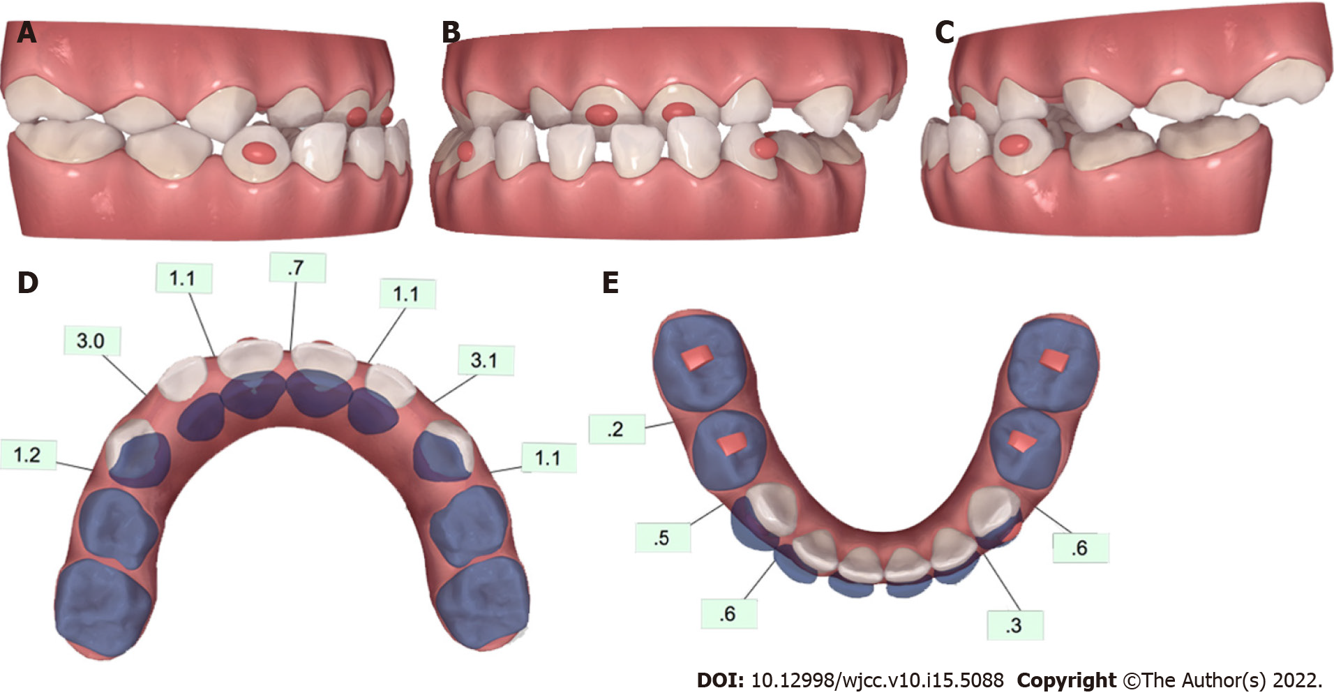
Figure 2 Pre-treatment ClinCheck® models and ClinCheck® treatment plan.
A: Lateral view of right side of pre-treatment model; B: Frontal view of pre-treatment model; C: Lateral view of left side of pre-treatment model; D: Occlusal view of superimposition of pre-treatment (blue) and post-treatment (white) maxillary arch models; E: Occlusal view of superimposition of pre-treatment (blue) and post-treatment (white) mandibular arch models.
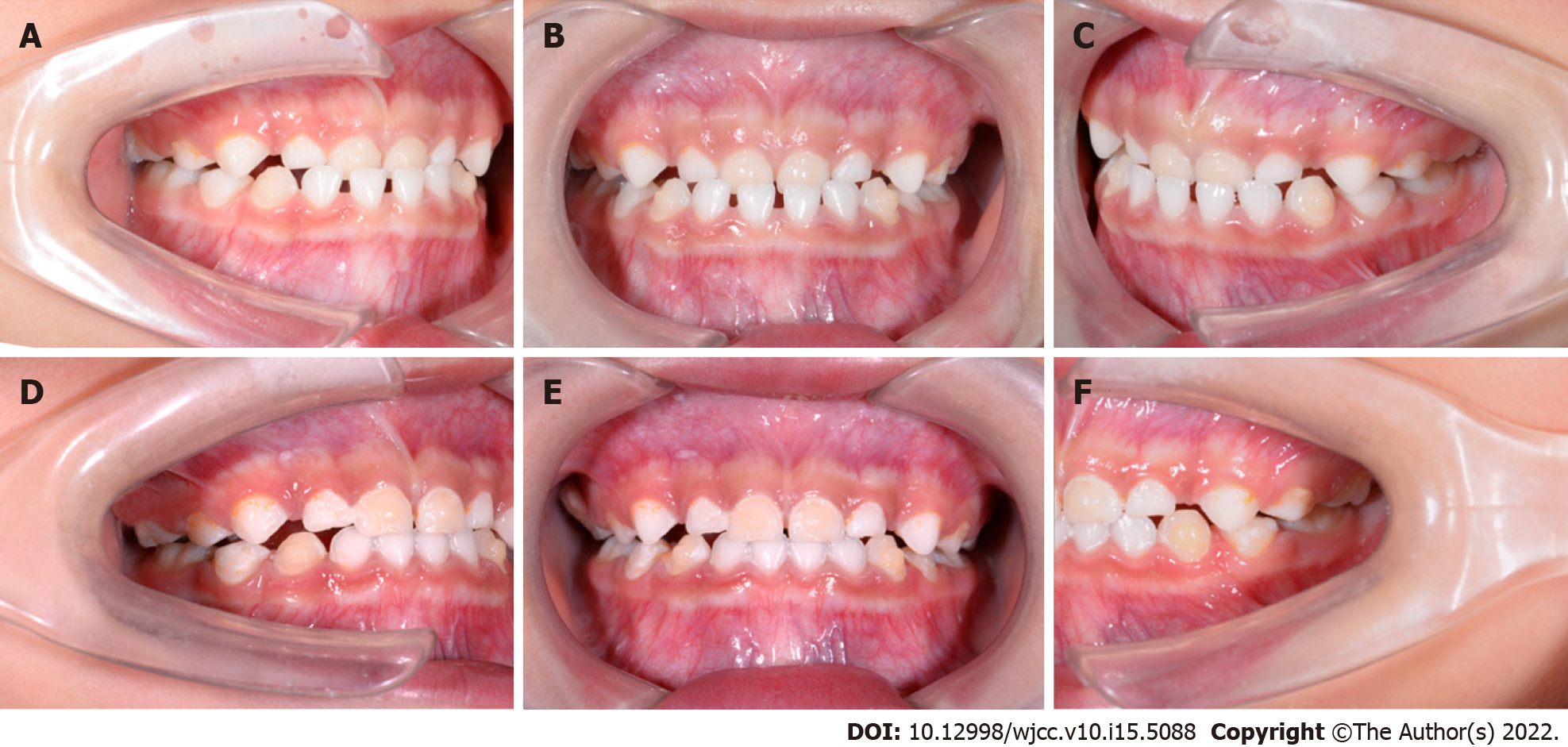
Figure 3 Intraoral photographs under treatment.
A: Lateral view of right side of intraoral pictures of the 4th aligner; B: Frontal view of intraoral pictures of the 4th aligner; C: Lateral view of left side of intraoral pictures of the 4th aligner; D: Lateral view of right side of intraoral pictures of the 13th aligner; E: Intraoral pictures of the 13th aligner; F: Lateral view of left side of intraoral pictures of the 13th aligner.
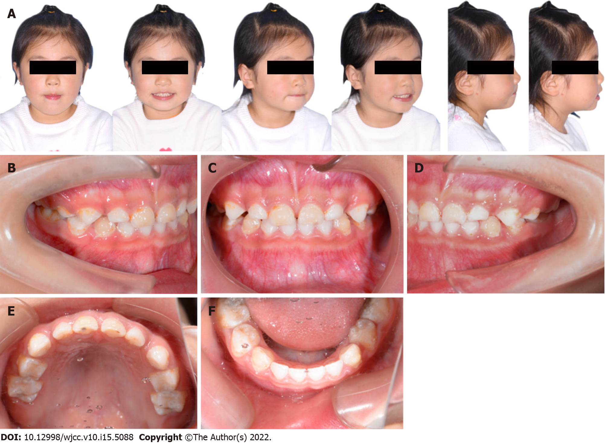
Figure 4 Post-treatment facial and intraoral photographs of the 19th aligner.
A: Post-treatment facial photos showing correction of facial asymmetry and harmonic appearance; B: Lateral view of right side; C: Frontal view showing correction of anterior cross-bite; D: Lateral view of left side; E: Occlusal view of the mandibular arch; F: Occlusal view of the maxillary arch.
Figure 5 Intraoral photographs of a 6-month follow-up.
A: Lateral view of right side; B: Frontal view; C: Lateral view of left side.
Figure 6 Facial and intraoral photographs of a 3-year follow-up.
A: 3-year follow-up facial photos; B: Lateral view of right side; C: Frontal view; D: Lateral view of left side; E: Occlusal view of the mandibular arch; F: Occlusal view of the maxillary arch.
- Citation: Zou YR, Gan ZQ, Zhao LX. Clear aligner treatment for a four-year-old patient with anterior cross-bite and facial asymmetry: A case report. World J Clin Cases 2022; 10(15): 5088-5096
- URL: https://www.wjgnet.com/2307-8960/full/v10/i15/5088.htm
- DOI: https://dx.doi.org/10.12998/wjcc.v10.i15.5088


