Copyright
©The Author(s) 2022.
World J Clin Cases. May 26, 2022; 10(15): 5072-5076
Published online May 26, 2022. doi: 10.12998/wjcc.v10.i15.5072
Published online May 26, 2022. doi: 10.12998/wjcc.v10.i15.5072
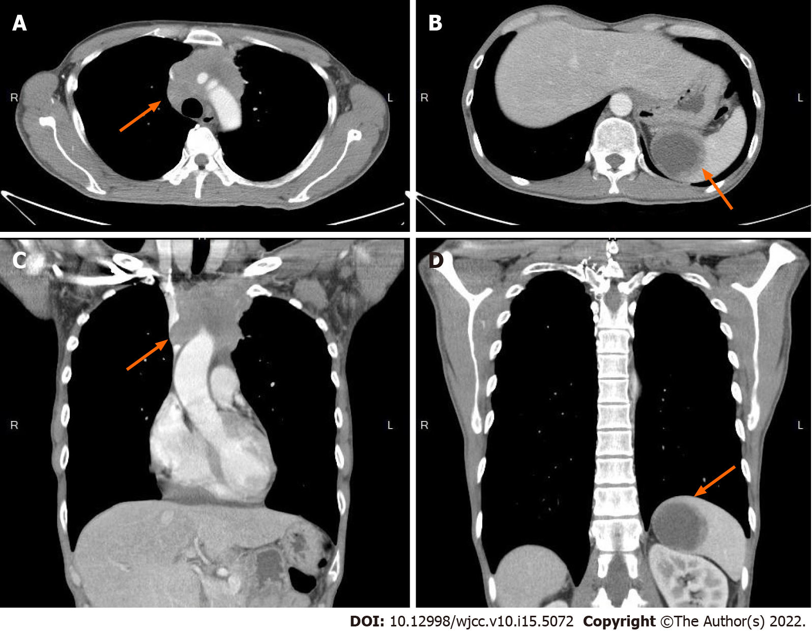
Figure 1 Computed tomography revealed mass over mediastinum and spleen.
A and C: The mediastinum has a confluent and lobulated soft tissue mass (arrow), encasing the right brachiocephalic artery, right and left carotid arteries, and left subclavian artery and compressing the superior vena cava; B and D: There is one cystic mass (arrow), measuring approximately 5.1-cm in diameter, in the spleen.
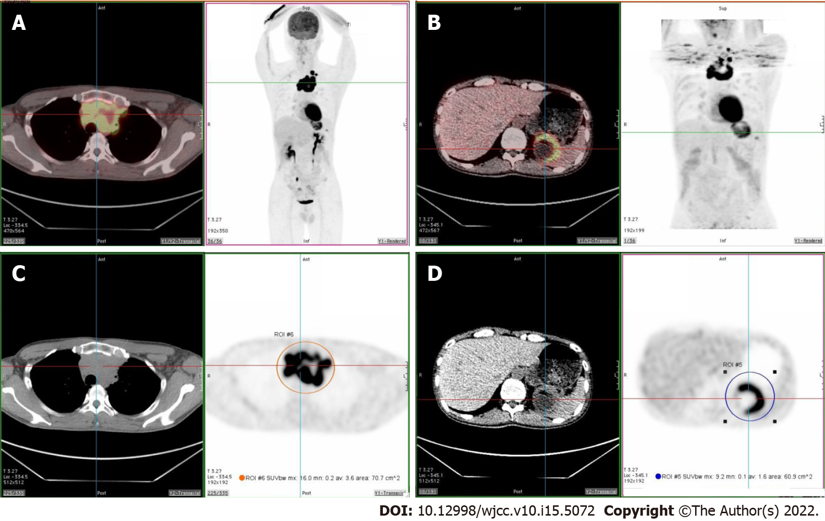
Figure 2 Fluorine-18 fluorodeoxyglucose positron emission tomography/computed tomography revealed increased fluorodeoxyglucose uptake over superior mediastinum and spleen.
A and C: Intense fluorodeoxyglucose (FDG) avidity in a soft tissue lobulated mass occupying the superior mediastinum (SUVmax = 16.0, size > 9.0 cm); B and D: Intense FDG avidity over the cystic lesion in the spleen (SUVmax = 9.2, size = 6.1 cm).
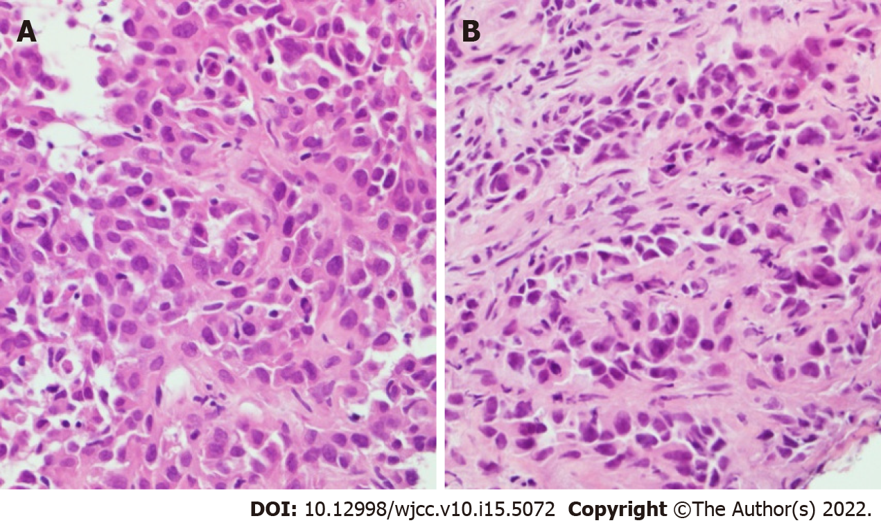
Figure 3 Pathology images.
A: Hematoxylin-Eosin staining of the thymic mass showed polygonal and spindle shaped tumor cells accompanied by giant cells with bizarre and multilobulated nuclei; B: Hematoxylin-Eosin staining of the spleen tissue was compatible with poorly differentiated metastatic carcinoma.
- Citation: Tsai YH, Lin KH, Huang TW. Rare solitary splenic metastasis from a thymic carcinoma detected on fluorodeoxyglucose-positron emission tomography: A case report. World J Clin Cases 2022; 10(15): 5072-5076
- URL: https://www.wjgnet.com/2307-8960/full/v10/i15/5072.htm
- DOI: https://dx.doi.org/10.12998/wjcc.v10.i15.5072


