Copyright
©The Author(s) 2022.
World J Clin Cases. May 26, 2022; 10(15): 5051-5056
Published online May 26, 2022. doi: 10.12998/wjcc.v10.i15.5051
Published online May 26, 2022. doi: 10.12998/wjcc.v10.i15.5051
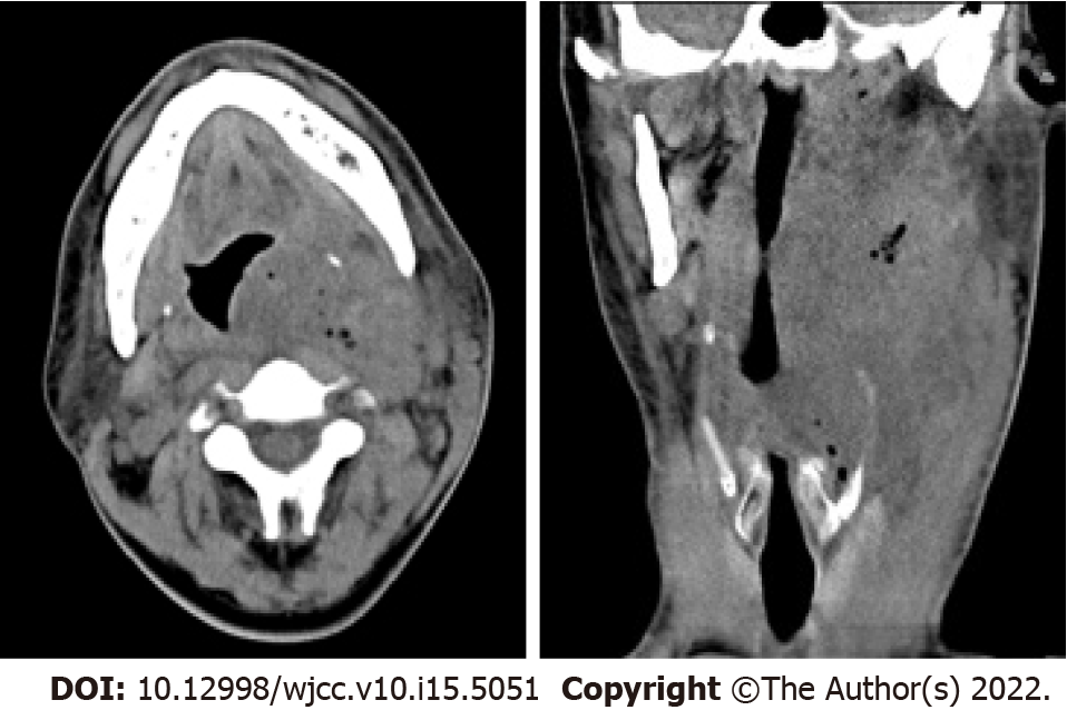
Figure 1 Plain computed tomography revealed a soft tissue mass containing scattered air (dimensions: 9.
9 cm × 7.3 cm × 4.6 cm; computed tomography value: 22-34 HU) that had a vague boundary and an irregular shape on the left neck and submaxillary space, and the oropharynx and laryngeal left lateral wall were thickened.
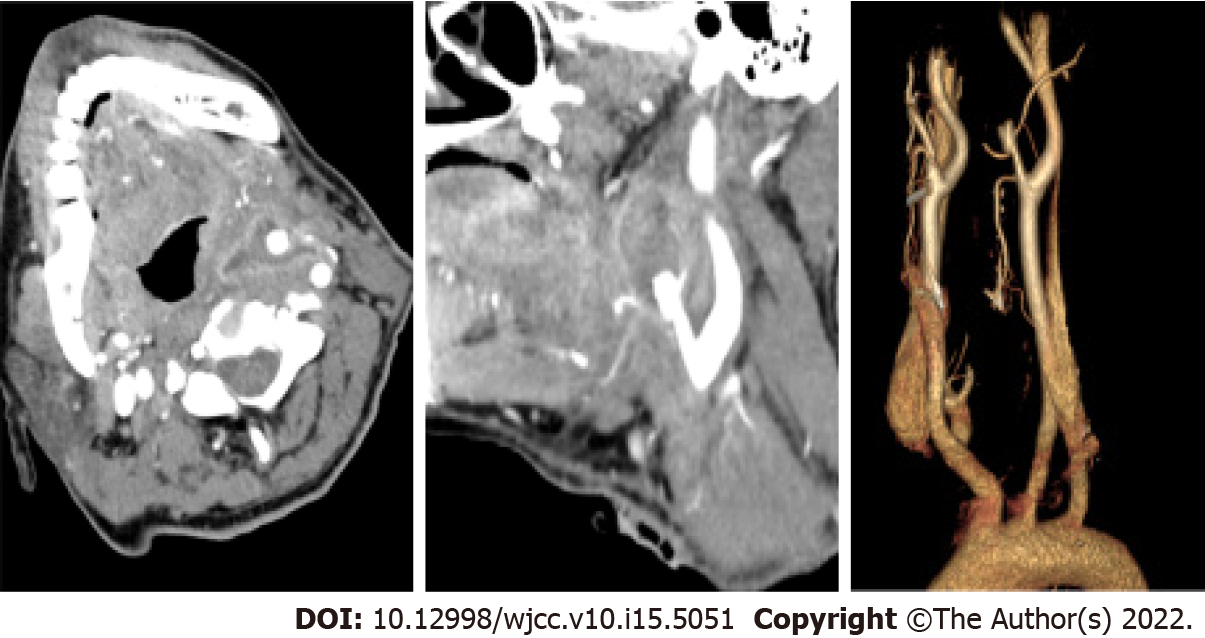
Figure 2 The enhanced computed tomography showed distal occlusion of the left external carotid artery and irregular thickening of the broken ends of the artery encased in an uneven enhancement of soft tissue density.
- Citation: Xie TH, Zhao WJ, Li XL, Hou Y, Wang X, Zhang J, An XH, Liu LT. Carotid blowout syndrome caused by chronic infection: A case report. World J Clin Cases 2022; 10(15): 5051-5056
- URL: https://www.wjgnet.com/2307-8960/full/v10/i15/5051.htm
- DOI: https://dx.doi.org/10.12998/wjcc.v10.i15.5051


