Copyright
©The Author(s) 2022.
World J Clin Cases. May 26, 2022; 10(15): 5036-5041
Published online May 26, 2022. doi: 10.12998/wjcc.v10.i15.5036
Published online May 26, 2022. doi: 10.12998/wjcc.v10.i15.5036
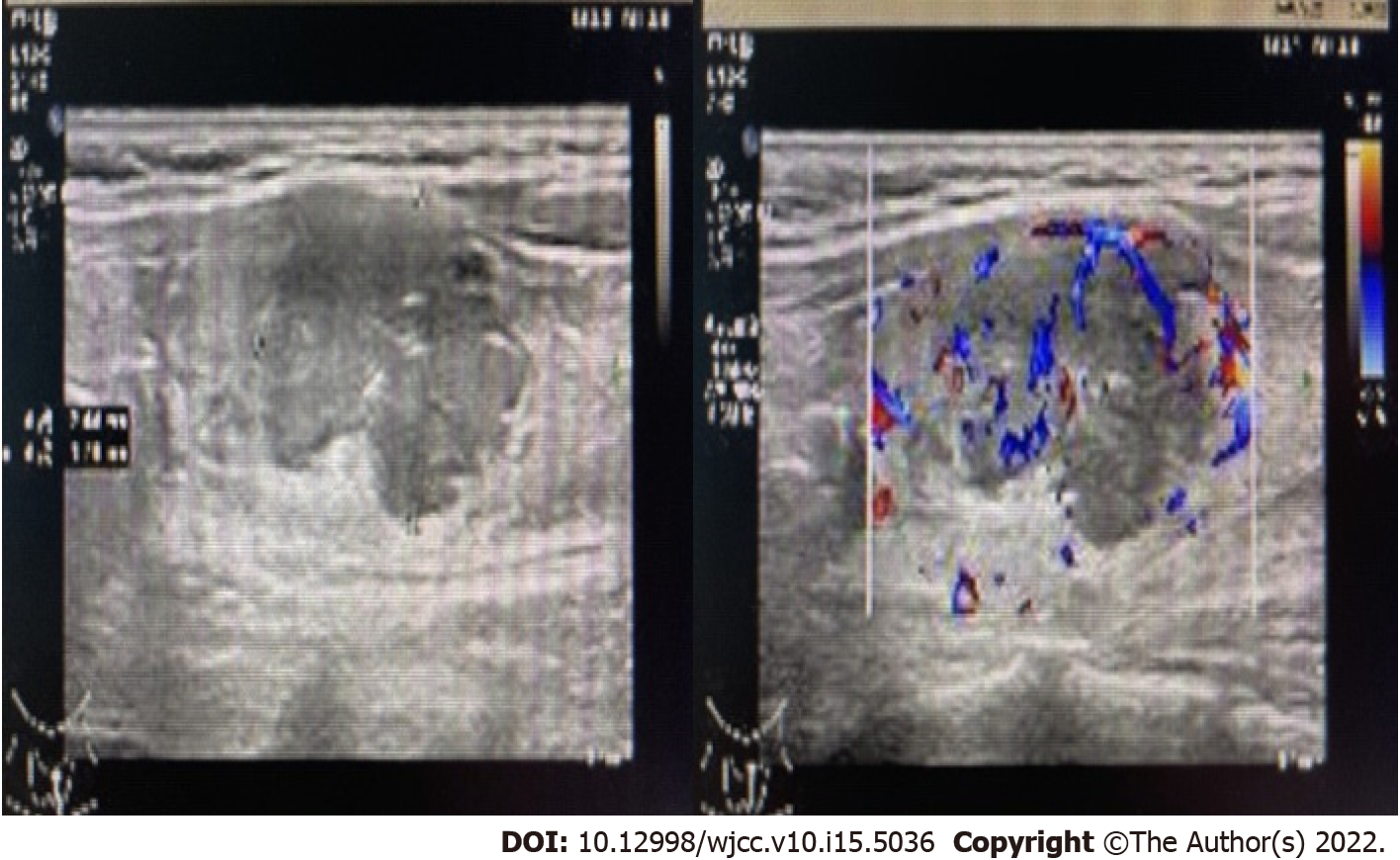
Figure 1 Thyroid color Doppler ultrasound showed hypoechoic nodules in the left lobe of the thyroid (TI-RADS 4b).
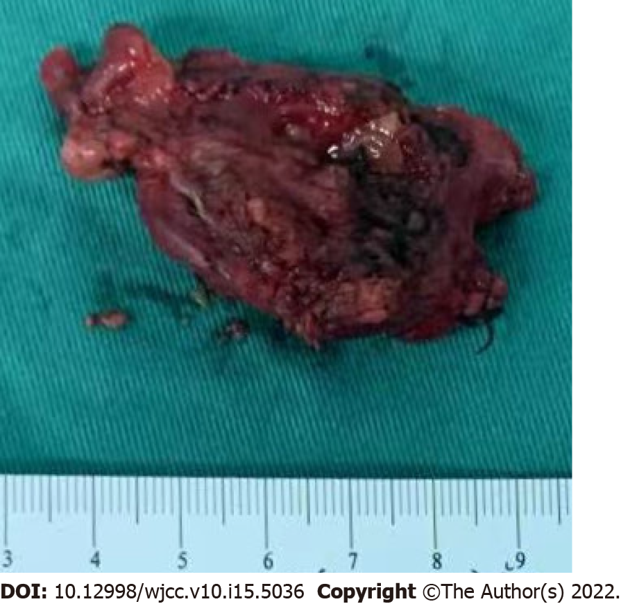
Figure 2 Tumor infiltration seen in the thyroid tissue.
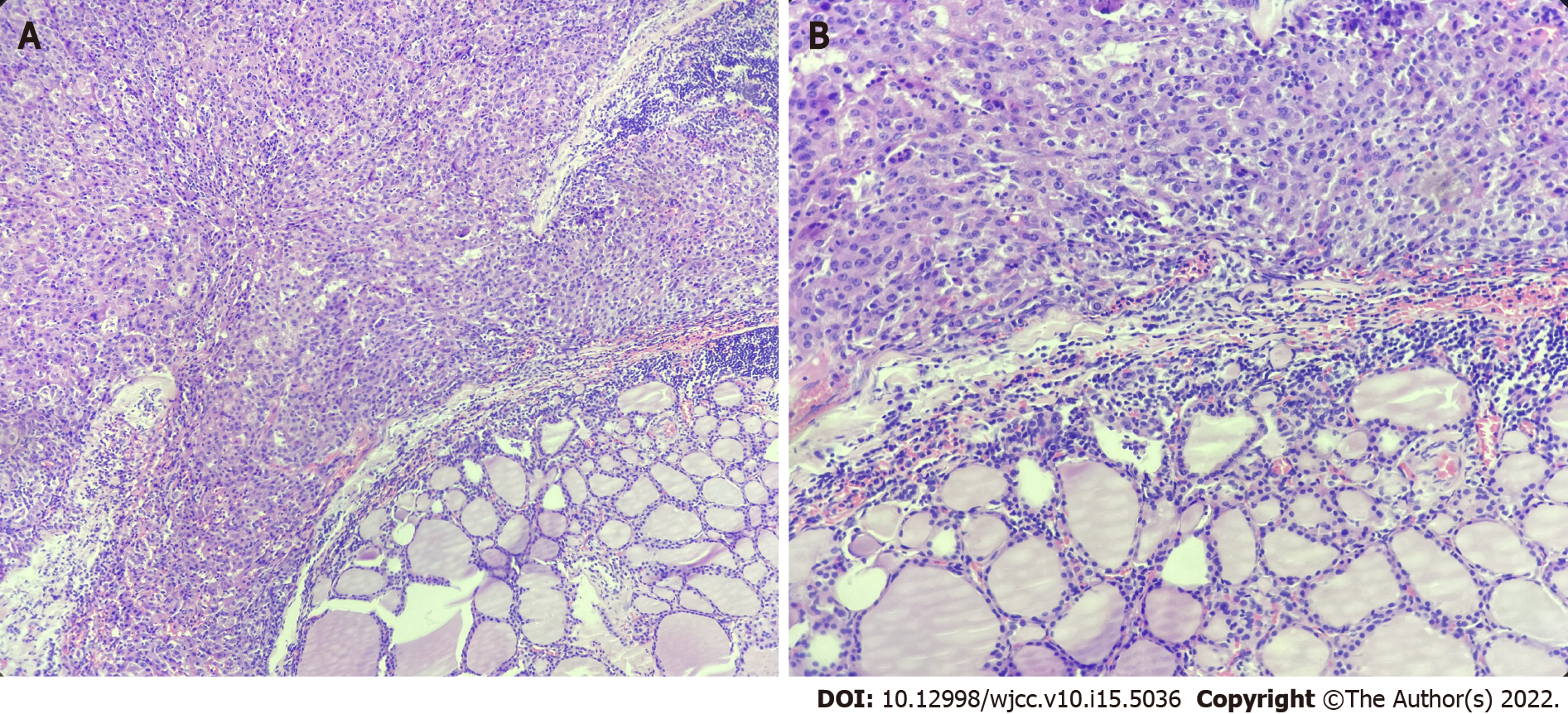
Figure 3 Pathology showed liver cancer infiltration in thyroid tissue.
A: H&E, × 100; B: H&E, × 200.
Figure 4 Immunohistochemical examination was positive for hepatocytes (× 100) and arginase-1 (× 100), and negative for TTF-1 (× 100).
A: Hepatocytes (× 100); B: Arginase-1 (× 100); C: TTF-1 (× 100).
- Citation: Zhong HC, Sun ZW, Cao GH, Zhao W, Ma K, Zhang BY, Feng YJ. Metastasis of liver cancer to the thyroid after surgery: A case report. World J Clin Cases 2022; 10(15): 5036-5041
- URL: https://www.wjgnet.com/2307-8960/full/v10/i15/5036.htm
- DOI: https://dx.doi.org/10.12998/wjcc.v10.i15.5036


