Copyright
©The Author(s) 2022.
World J Clin Cases. May 26, 2022; 10(15): 4985-4990
Published online May 26, 2022. doi: 10.12998/wjcc.v10.i15.4985
Published online May 26, 2022. doi: 10.12998/wjcc.v10.i15.4985
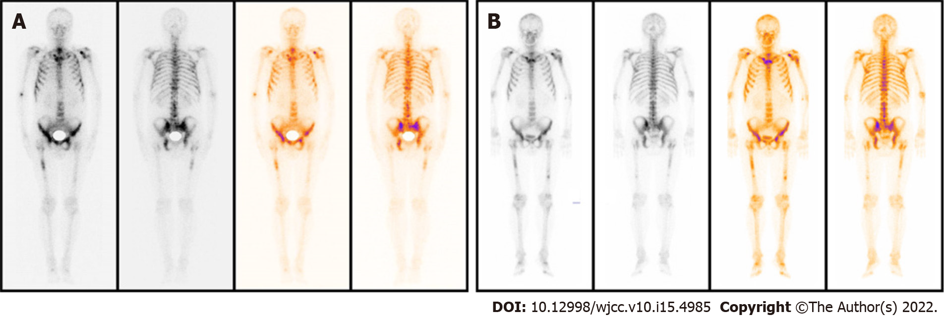
Figure 1 Radionuclide bone imaging indicated progression on bone.
A: Before the treatment; B: Three months later.
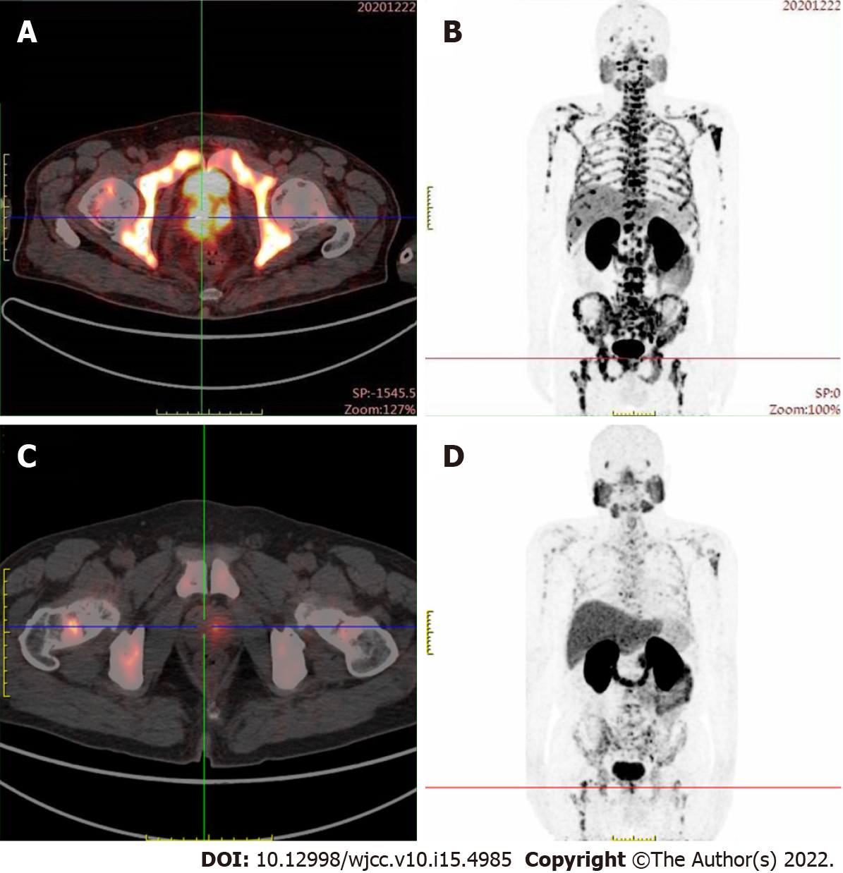
Figure 2 Prostate-specific membrane antigen positron emission tomography – computed tomography examination showed abnormal prostate-specific membrane antigen aggregation was significantly reduced, and bone metastasis was significantly improved compared with baseline.
A, B: Before the treatment; C, D: Three months later.
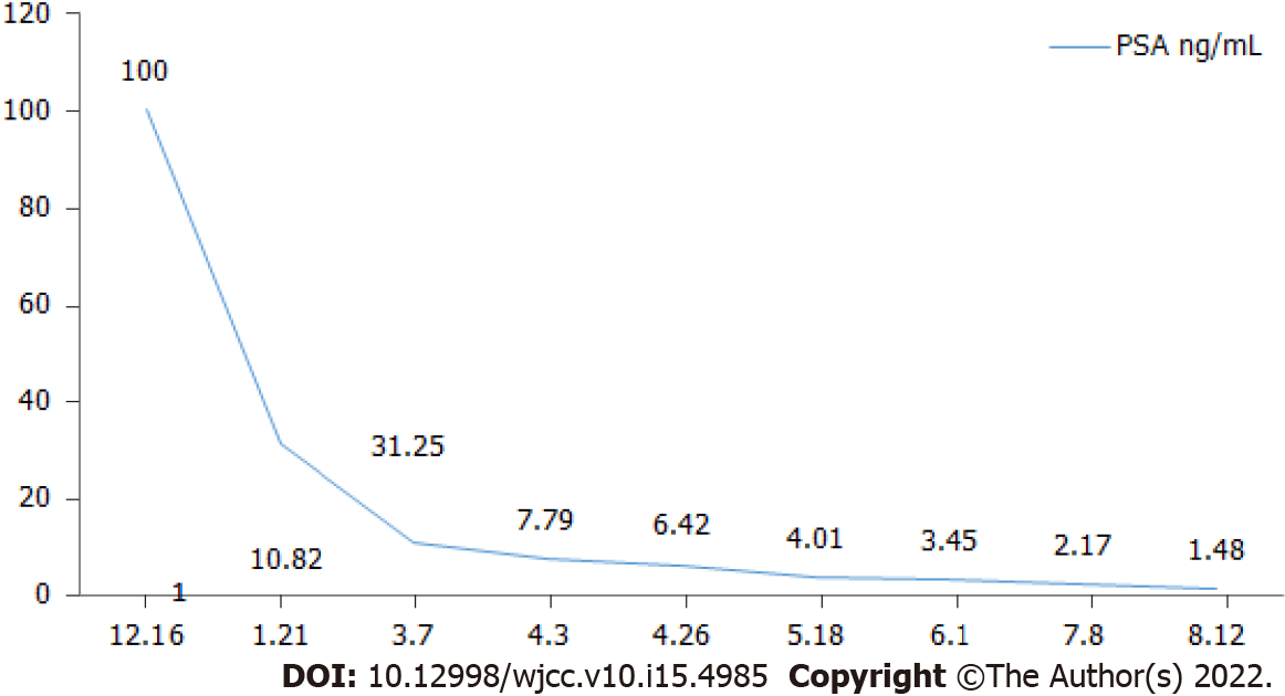
Figure 3 Prostate-specific antigen curve.
- Citation: Li KH, Du YC, Yang DY, Yu XY, Zhang XP, Li YX, Qiao L. Bone flare after initiation of novel hormonal therapy in patients with metastatic hormone-sensitive prostate cancer: A case report. World J Clin Cases 2022; 10(15): 4985-4990
- URL: https://www.wjgnet.com/2307-8960/full/v10/i15/4985.htm
- DOI: https://dx.doi.org/10.12998/wjcc.v10.i15.4985


