Copyright
©The Author(s) 2022.
World J Clin Cases. May 26, 2022; 10(15): 4929-4934
Published online May 26, 2022. doi: 10.12998/wjcc.v10.i15.4929
Published online May 26, 2022. doi: 10.12998/wjcc.v10.i15.4929
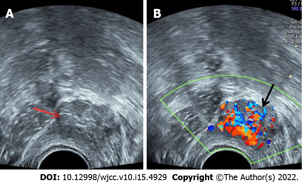
Figure 1 Transvaginal ultrasound showed a 2.
5 cm × 2.1 cm medium-echo mass protruding from the right anterior wall of the bladder (A, indicated by the red arrow); a regular shape, a clear boundary, a wide base, and rich strip blood flow signals were detected (B, indicated by the black arrow).
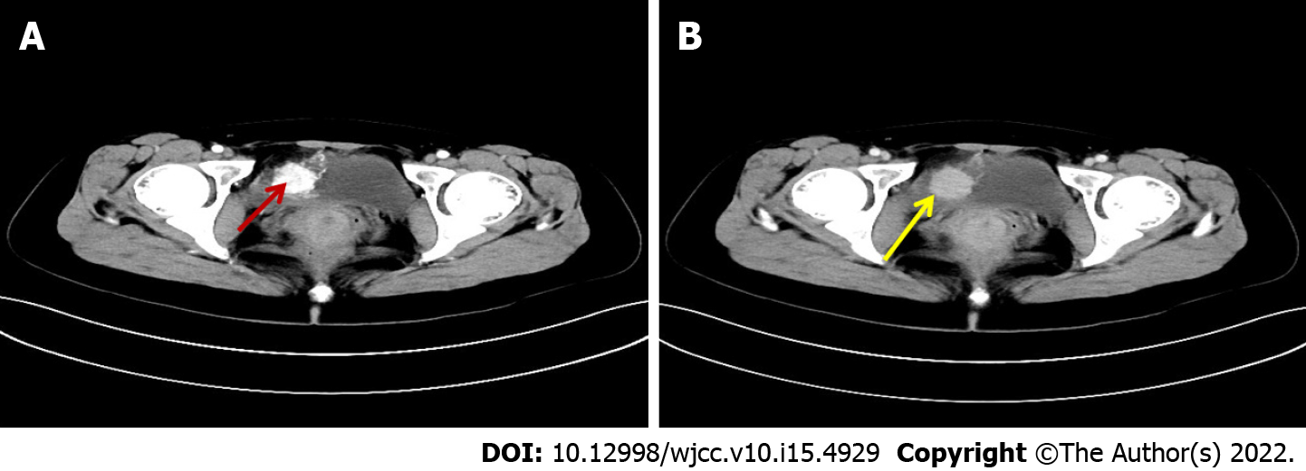
Figure 2 Enhanced computed tomography scan of the bladder showed a circular space-occupying lesion on the right anterior wall of the bladder, with obvious enhancement in the arterial phase (A, indicated by the red arrow) and weakening of the enhancement in the delayed phase (B, indicated by the yellow arrow).
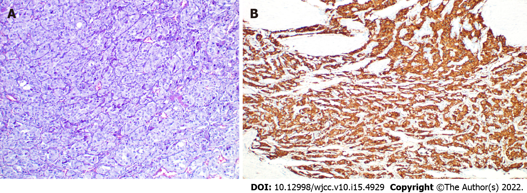
Figure 3 Pathological and immunological features of a paraganglioma of the urinary bladder.
A: Micrograph of conventional HE staining (× 200) showing chief cells in a nested growth pattern; B: The image shows positive expression of CgA in immunohistochemistry.
- Citation: Chen J, Yang HF. Nonfunctional bladder paraganglioma misdiagnosed as hemangioma: A case report. World J Clin Cases 2022; 10(15): 4929-4934
- URL: https://www.wjgnet.com/2307-8960/full/v10/i15/4929.htm
- DOI: https://dx.doi.org/10.12998/wjcc.v10.i15.4929


