Copyright
©The Author(s) 2022.
World J Clin Cases. May 26, 2022; 10(15): 4904-4910
Published online May 26, 2022. doi: 10.12998/wjcc.v10.i15.4904
Published online May 26, 2022. doi: 10.12998/wjcc.v10.i15.4904
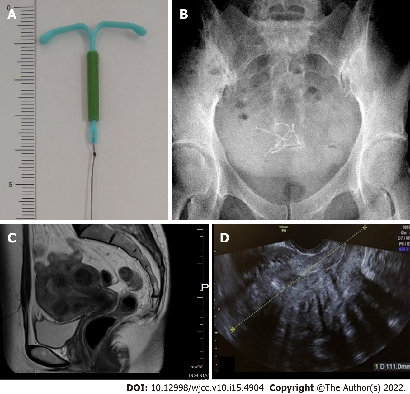
Figure 1 Photographic and imaging findings of levonorgestrel-releasing intrauterine system.
A: The T frame and thread are made of polyethylene, and barium sulfate is added to the T frame. The vertical portion of the cylinder contains Levonorgestrel-releasing intrauterine system (LNG-IUS); B: Abdominopelvic radiograph demonstrates the two LNG-IUSs in the pelvis and that both devices are rotated away from a normal orientation; C: Axial T2-weighted MR image through the pelvis before LNG-IUS insertion. Multiple fibroids are observed in the mucosa and muscle layer; D: Transvaginal ultrasonography at the first visit to our hospital. At this time, two LNG-IUSs were inserted, but due to multiple fibroids, it was difficult to confirm the device location by ultrasound.
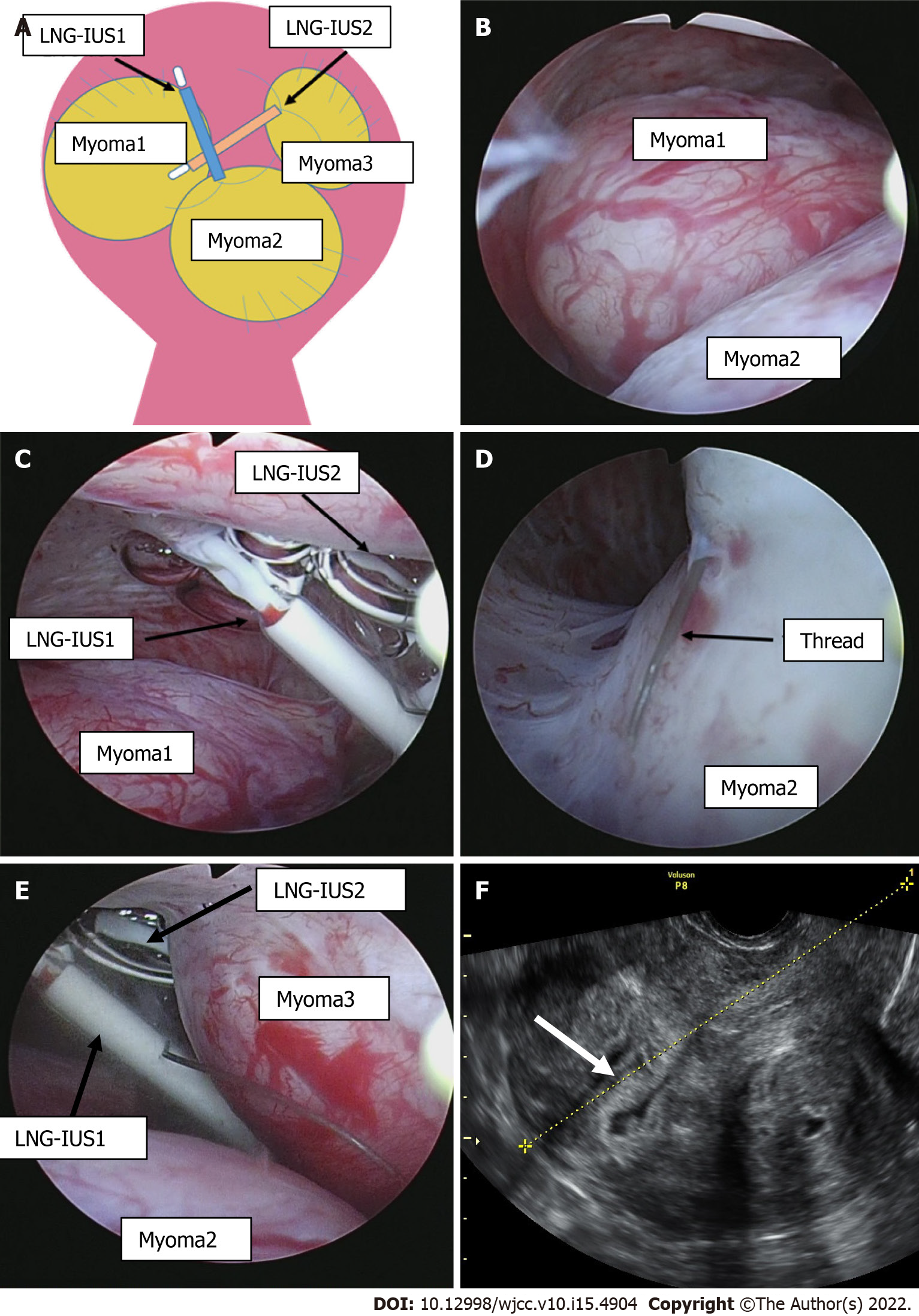
Figure 2 Hysteroscopic findings in the endometrial cavity and ultrasound findings.
A: Illustration of the uterus, fibroids and devices; B: Hysteroscopic findings show multiple fibroids occupying the uterus; C: Levonorgestrel-releasing intrauterine system (LNG-IUS) had perforated the myometrium of the anterior wall of the uterine body; D: A thread had penetrated into the muscle layer; E: LNG-IUS1 and 2 were further toward the bottom of the uterus than myoma 2 and 3; F: The endometrium was smooth on transvaginal ultrasound (white arrow).
- Citation: Maebayashi A, Kato K, Hayashi N, Nagaishi M, Kawana K. Importance of abdominal X-ray to confirm the position of levonorgestrel-releasing intrauterine system: A case report. World J Clin Cases 2022; 10(15): 4904-4910
- URL: https://www.wjgnet.com/2307-8960/full/v10/i15/4904.htm
- DOI: https://dx.doi.org/10.12998/wjcc.v10.i15.4904


