Copyright
©The Author(s) 2022.
World J Clin Cases. May 26, 2022; 10(15): 4776-4784
Published online May 26, 2022. doi: 10.12998/wjcc.v10.i15.4776
Published online May 26, 2022. doi: 10.12998/wjcc.v10.i15.4776
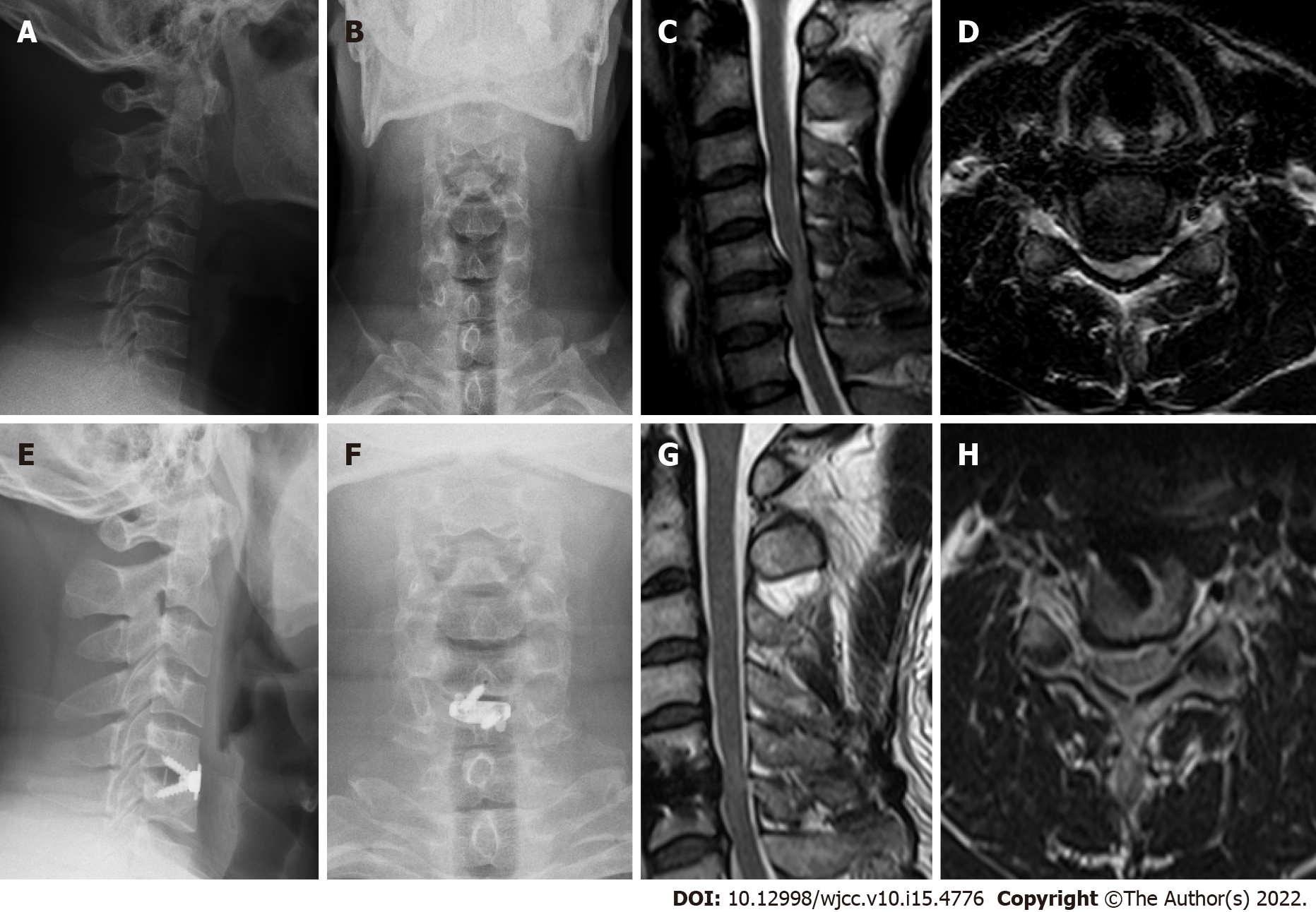
Figure 1 A 41-year-old male with spinal cord compression at C5/6 level was treated by anterior cervical discectomy and fusion with self-locking fusion cage.
The self-locking fusion cage was in good position, the decompression was sufficient, and the clinical symptoms significantly improved. A: Preoperative lateral X-ray; B: Preoperative anteroposterior X-ray; C: Preoperative lateral magnetic resonance imaging (MRI); D: Preoperative axial MRI; E: Lateral X-ray at last follow-up; F: Anteroposterior X-ray at last follow-up; G: Lateral MRI at last follow-up; H: Axial MRI at last follow-up.
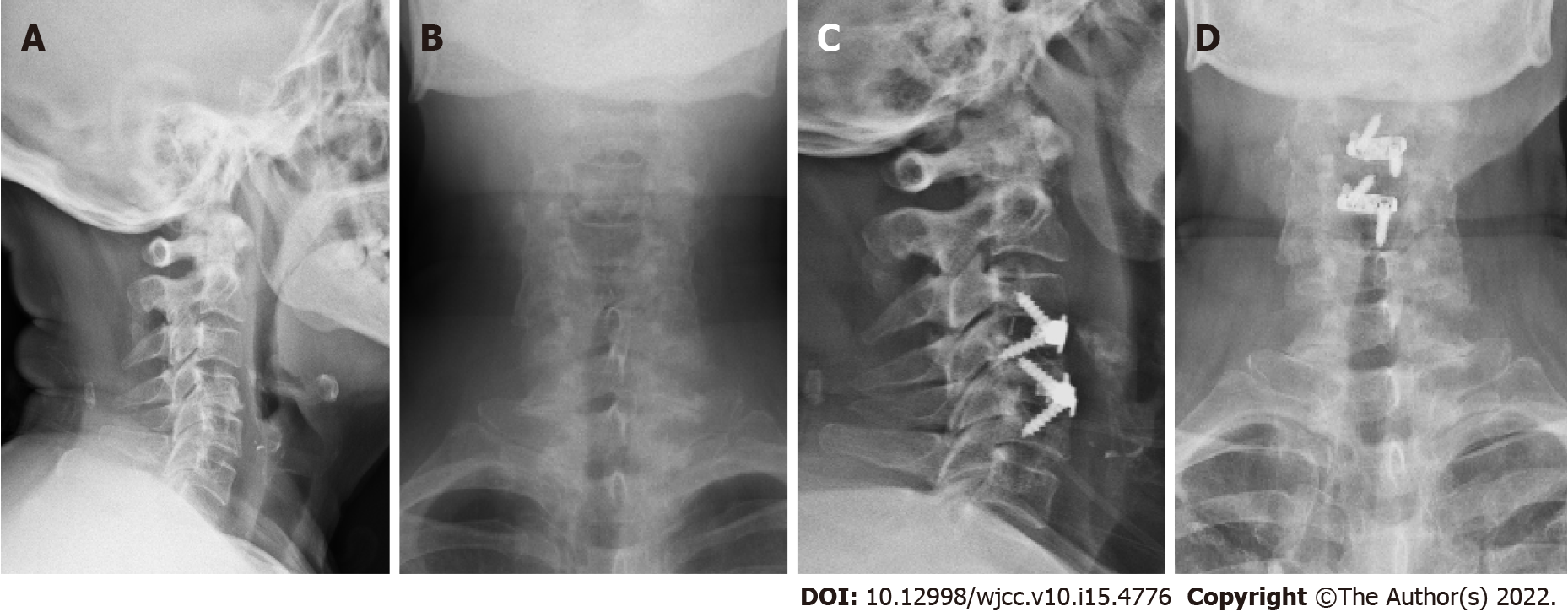
Figure 2 A 76-year-old female was treated by bi-level anterior cervical discectomy and fusion with self-locking fusion cage at C3/4 and C4/5 level.
A: Preoperative lateral X-ray; B: Preoperative anteroposterior X-ray; C: Lateral X-ray at last follow-up; D: Anteroposterior X-ray at last follow-up.
- Citation: Zhang B, Jiang YZ, Song QP, An Y. Outcomes of cervical degenerative disc disease treated by anterior cervical discectomy and fusion with self-locking fusion cage. World J Clin Cases 2022; 10(15): 4776-4784
- URL: https://www.wjgnet.com/2307-8960/full/v10/i15/4776.htm
- DOI: https://dx.doi.org/10.12998/wjcc.v10.i15.4776


