Copyright
©The Author(s) 2022.
World J Clin Cases. May 16, 2022; 10(14): 4684-4690
Published online May 16, 2022. doi: 10.12998/wjcc.v10.i14.4684
Published online May 16, 2022. doi: 10.12998/wjcc.v10.i14.4684
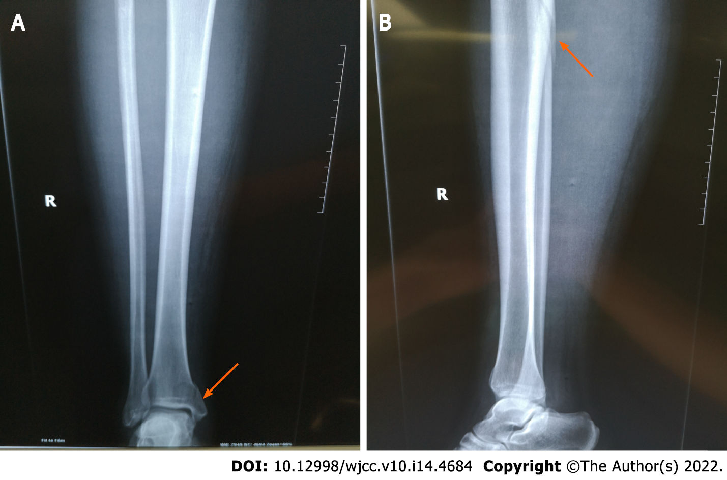
Figure 1 Preoperative radiographic view of the ankle and leg.
A: Preoperative anteroposterior radiographic view of the ankle and leg (arrow); B: Preoperative lateral radiographic view of the ankle and leg (arrow). Full-length radiographs of the lower leg revealed proximal fibula fracture (arrow) and medial malleolar fracture (arrow).
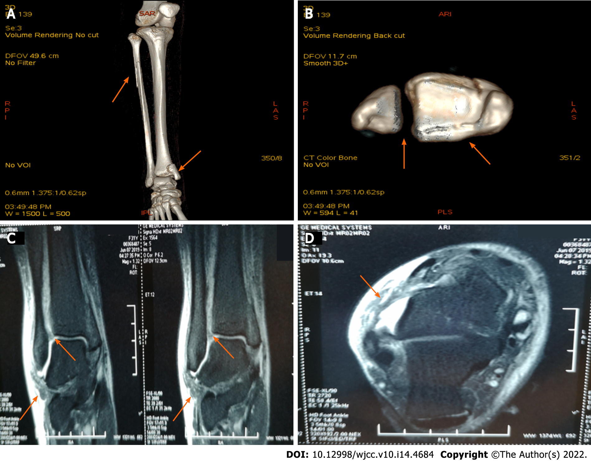
Figure 2 Three-dimensional computed tomography scan and magnetic resonance imaging of the ankle and leg.
A: Anterior view of the computed tomography (CT) scan of the ankle and leg (arrow); B: Axis view of the CT scan of the ankle (arrow); C: Sagittal magnetic resonance imaging (MRI) of the ankle (arrow); D: Axial MRI of the ankle (arrow); A and B: CT scan revealed proximal fibula fracture, inferior tibiofibular joint separation, and medial malleolar fracture involving the posterior malleolus (arrow); C and D: MRI revealed rupture of the anterior inferior tibiofibular ligament and anterior talofibular ligament (arrow).
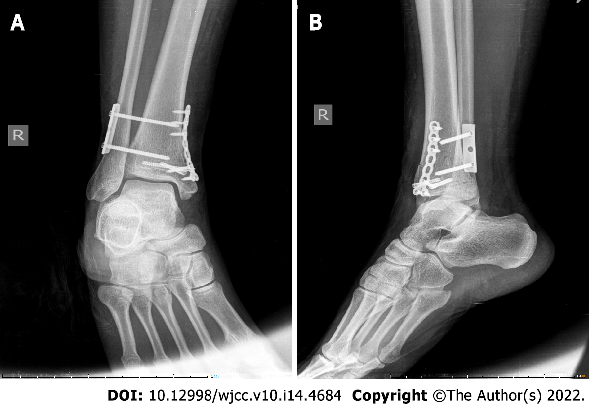
Figure 3 Immediate postoperative radiographic view of the ankle.
A: Immediate postoperative anteroposterior radiographic view of the ankle; B: Immediate postoperative lateral radiographic view of the ankle.
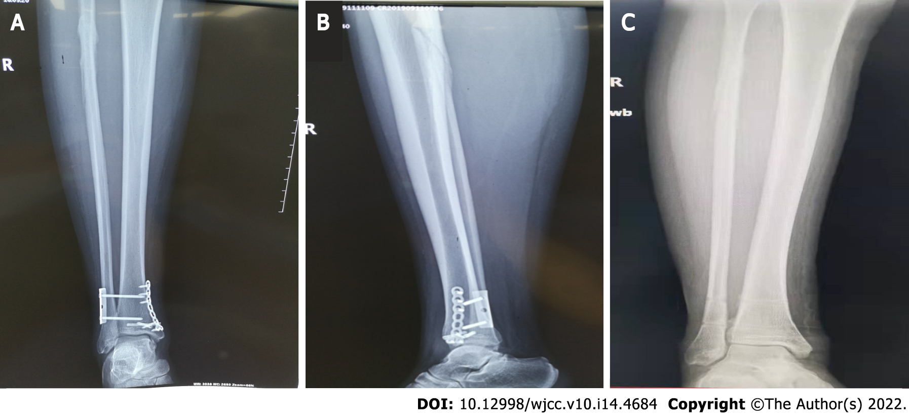
Figure 4 Three months postoperative radiographs of the ankle and leg.
A and B: Anteroposterior and lateral radiographic view of the ankle and leg before removal of internal fixation; C: Anteroposterior view of the ankle and leg after removal of internal fixation.
- Citation: Zhao B, Li N, Cao HB, Wang GX, He JQ. Rare pattern of Maisonneuve fracture: A case report. World J Clin Cases 2022; 10(14): 4684-4690
- URL: https://www.wjgnet.com/2307-8960/full/v10/i14/4684.htm
- DOI: https://dx.doi.org/10.12998/wjcc.v10.i14.4684


